Copyright
©The Author(s) 2023.
World J Clin Cases. Jan 6, 2023; 11(1): 172-176
Published online Jan 6, 2023. doi: 10.12998/wjcc.v11.i1.172
Published online Jan 6, 2023. doi: 10.12998/wjcc.v11.i1.172
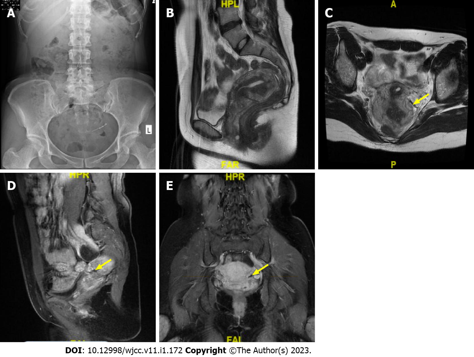
Figure 1 X-ray and magnetic resonance imaging of the malpositioned levonorgestrel-releasing intrauterine system.
A: X-ray shows that a radiopaque T-shape device is visible in the pelvis; B: The uterus is in the retroflexed position; C: The malpositioned levonorgestrel-releasing intrauterine system (yellow arrow) was seen in axial; D: Sagittal; E: Coronary position.
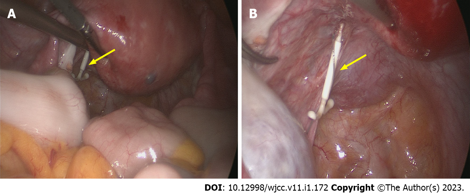
Figure 2 During the operation, an intact levonorgestrel-releasing intrauterine system with a tail wire was found near the left uterosacral ligament.
A: During the operation, the uterus is in the retroflexed position and an intact levonorgestrel-releasing intrauterine system with a tail wire is found near the left uterosacral ligament (A, yellow arrow); B: Without adhesion to the surrounding tissues (B, yellow arrow).
- Citation: Zhang GR, Yu X. Perforation of levonorgestrel-releasing intrauterine system found at one month after insertion: A case report. World J Clin Cases 2023; 11(1): 172-176
- URL: https://www.wjgnet.com/2307-8960/full/v11/i1/172.htm
- DOI: https://dx.doi.org/10.12998/wjcc.v11.i1.172


