Copyright
©The Author(s) 2023.
World J Clin Cases. Jan 6, 2023; 11(1): 164-171
Published online Jan 6, 2023. doi: 10.12998/wjcc.v11.i1.164
Published online Jan 6, 2023. doi: 10.12998/wjcc.v11.i1.164
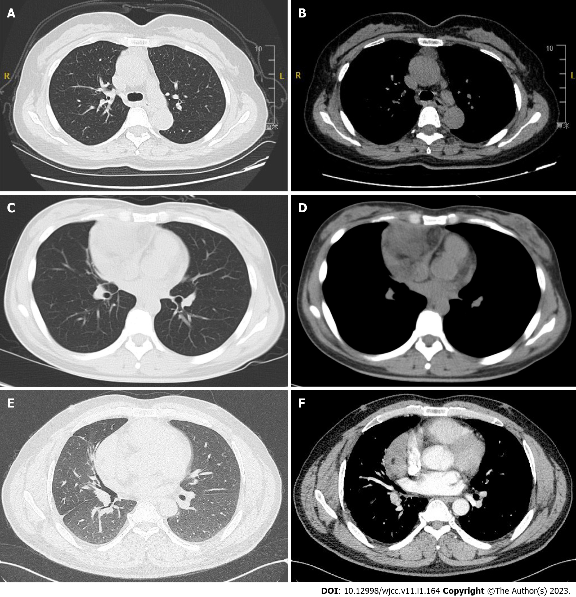
Figure 1 Computed tomography scan of the chest.
A and B: In case 1, computed tomography scan revealed the mass shadow in the anterior mediastinum, about 3.9 cm × 1.8 cm in size, with clear boundary; C and D: In case 2, computed tomography scan revealed irregular mass shadow in the anterior upper mediastinum, about 7.1 cm × 7.5 cm in size, with clear boundary, protruding to the right lung, uneven internal density, fat density shadow could be seen, and the boundary between the lesion and adjacent large blood vessels and cardiac structures was unclear; E and F: In case 3, computed tomography scan revealed lumpy shadows about 9.9 cm × 9.8 cm × 4.9 cm in size beside the anterior middle mediastinum, with spot-like calcification, and small patchy low-density shadows in it. The boundary of tumor mass was clear.
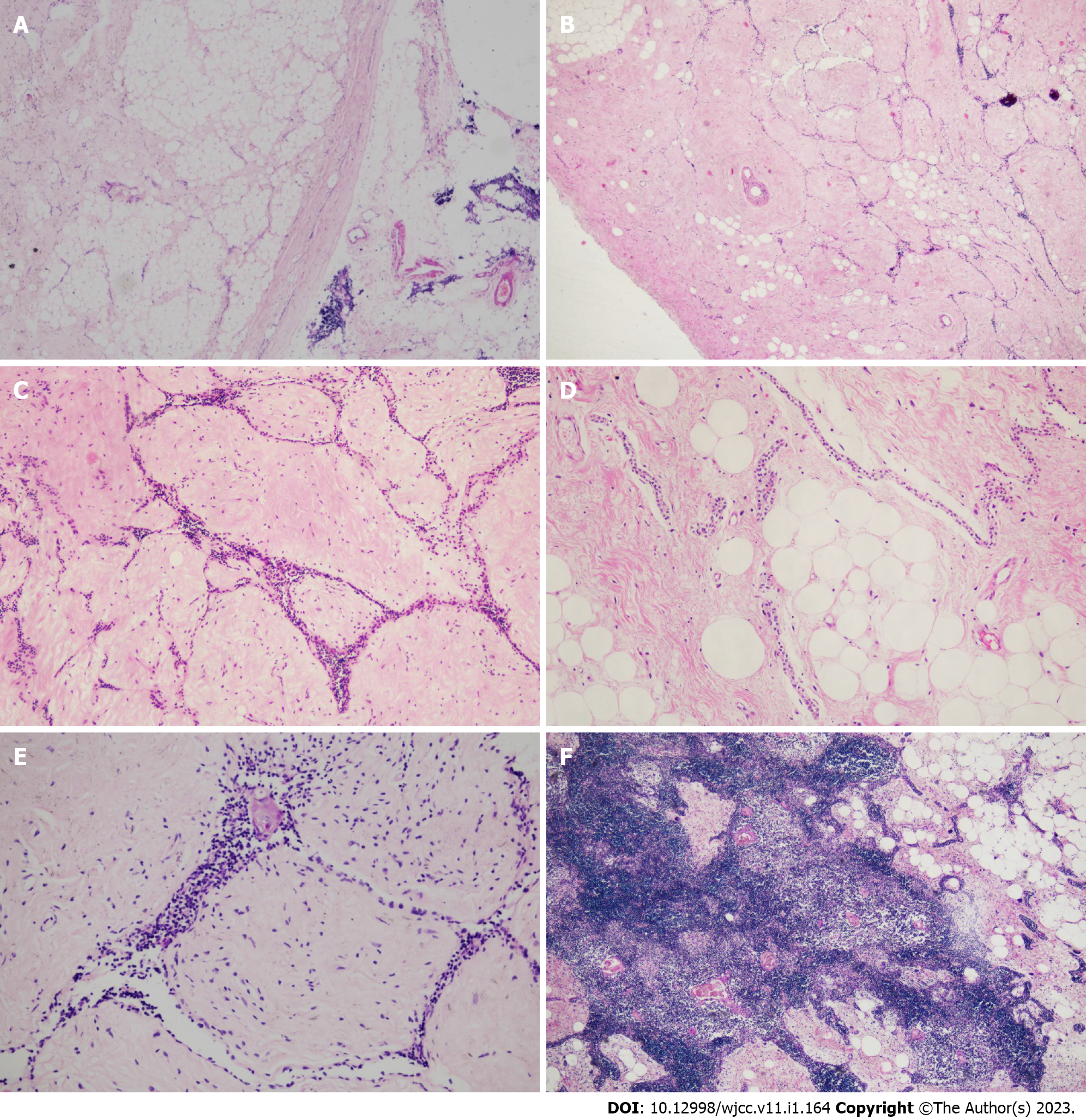
Figure 2 Histological features of the thymic lipofibroadenoma.
A: The tumor was all well circumscribed, and a clear connective capsule was observed between the tumor and the remaining thymus. [hematoxylin and eosin (H&E) staining, × 40]; B and C: The tumor was composed of fibrotic and hyaline stroma, narrow strands of epithelial cells and adipose tissues. The blank-looking epithelial cells formed narrow cords and interspersed in fibrotic and hyaline stroma. (H&E staining, B, × 40; C, × 100); D: Some adipocytes were mixed with the fibrotic stroma. Abundant adipose tissue also can be seen in some areas. (H&E staining, × 100); E: A few lymphocytes were mixed with the epithelial cells. Residual Hassall corpuscles and small calcifications could be seen in some region. (H&E staining, × 200); F: In case 2, hyperplastic thymic tissue was observed in part of the tumor. (H&E staining, × 100).
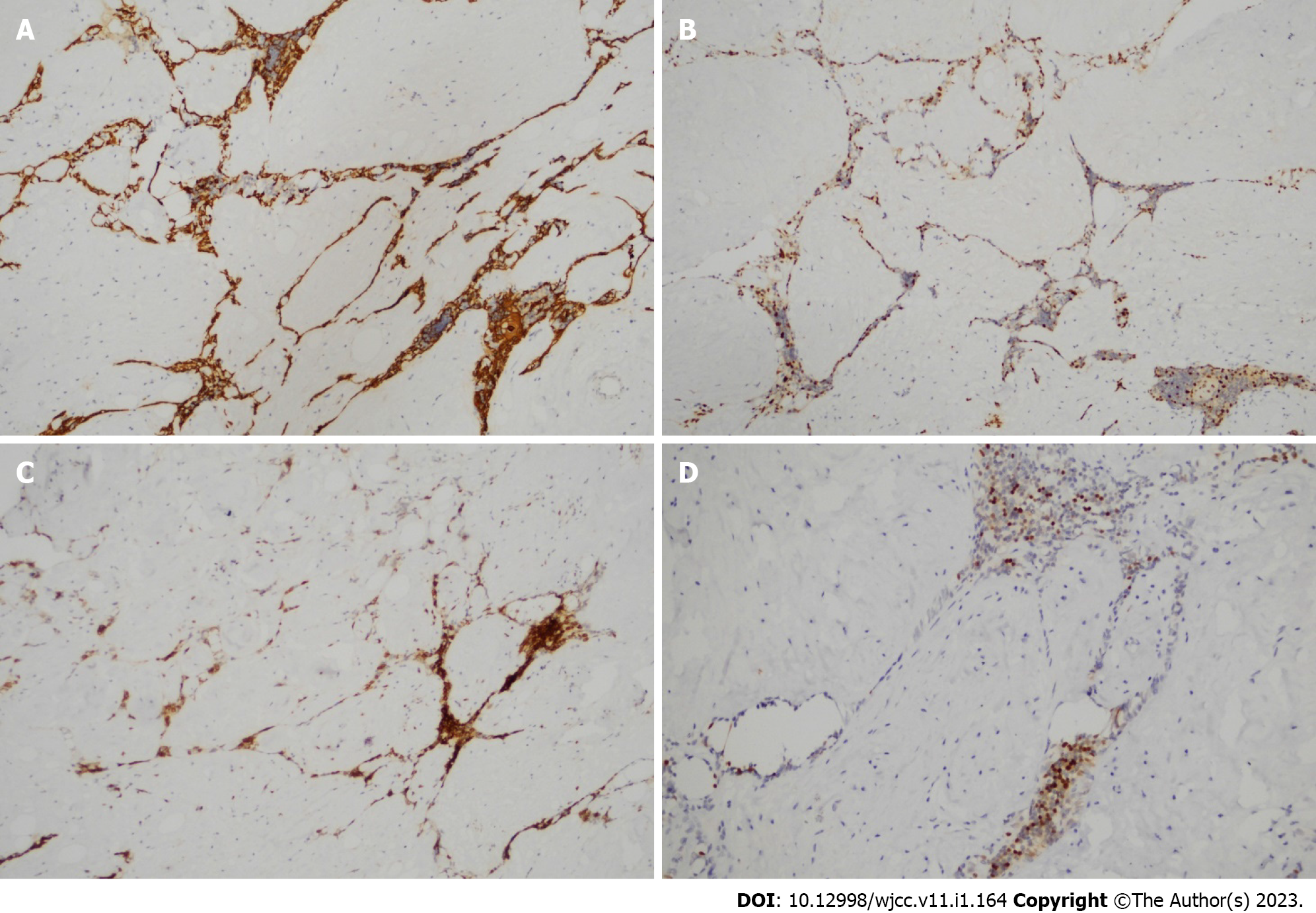
Figure 3 Immunohistochemical staining of thymic lipofibroadenoma.
A and B: Epithelial cells positive for broad-spectrum CK (A), and P63 (B) (× 100); C: lymphocytes were positive for CD3 (× 100); D: Some lymphocytes were positive for TdT (× 200).
- Citation: Yang MQ, Wang ZQ, Chen LQ, Gao SM, Fu XN, Zhang HN, Zhang KX, Xu HT. Thymic lipofibroadenomas: Three case reports. World J Clin Cases 2023; 11(1): 164-171
- URL: https://www.wjgnet.com/2307-8960/full/v11/i1/164.htm
- DOI: https://dx.doi.org/10.12998/wjcc.v11.i1.164


