Copyright
©The Author(s) 2023.
World J Clin Cases. Jan 6, 2023; 11(1): 157-163
Published online Jan 6, 2023. doi: 10.12998/wjcc.v11.i1.157
Published online Jan 6, 2023. doi: 10.12998/wjcc.v11.i1.157
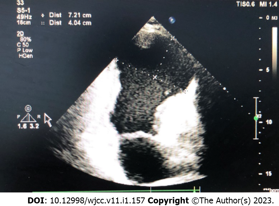
Figure 1 Preoperative echocardiographic image.
Left ventricular volume was significantly increased, and the formation of a huge ventricular aneurysm was seen at the apex.
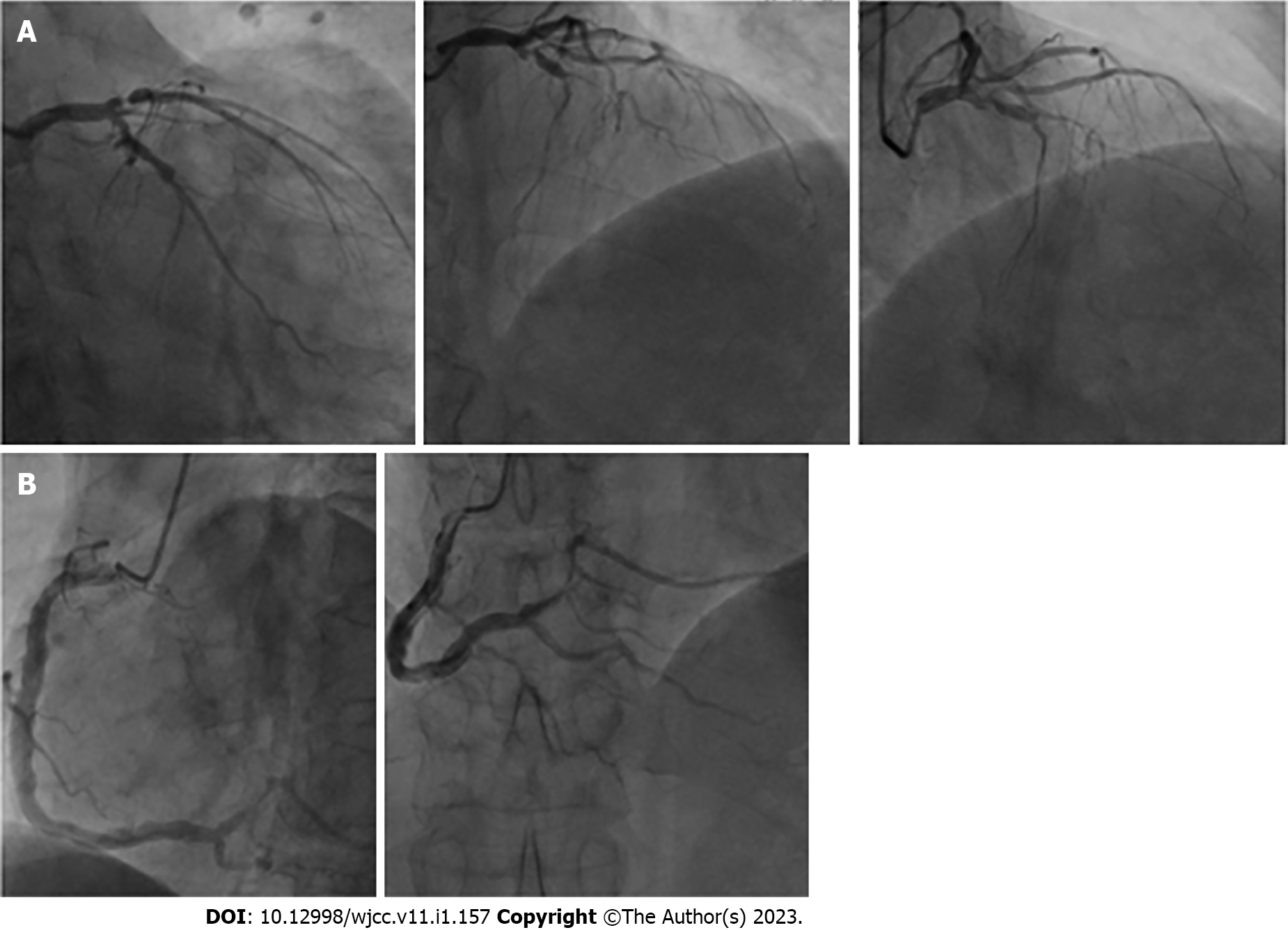
Figure 2 Coronary angiographic images.
A: Left coronary angiography showed 70%-80% stenosis in the proximal segment of the anterior descending artery, 100% stenosis in the proximal and middle segments, 70% stenosis in the proximal segment of D1, and 70% stenosis in the middle segment of the circumflex artery; B: Right coronary angiography aneurysm-like expansion, proximal 40%-50% stenosis, and 30%-40% stenosis in the distal segment were observed.
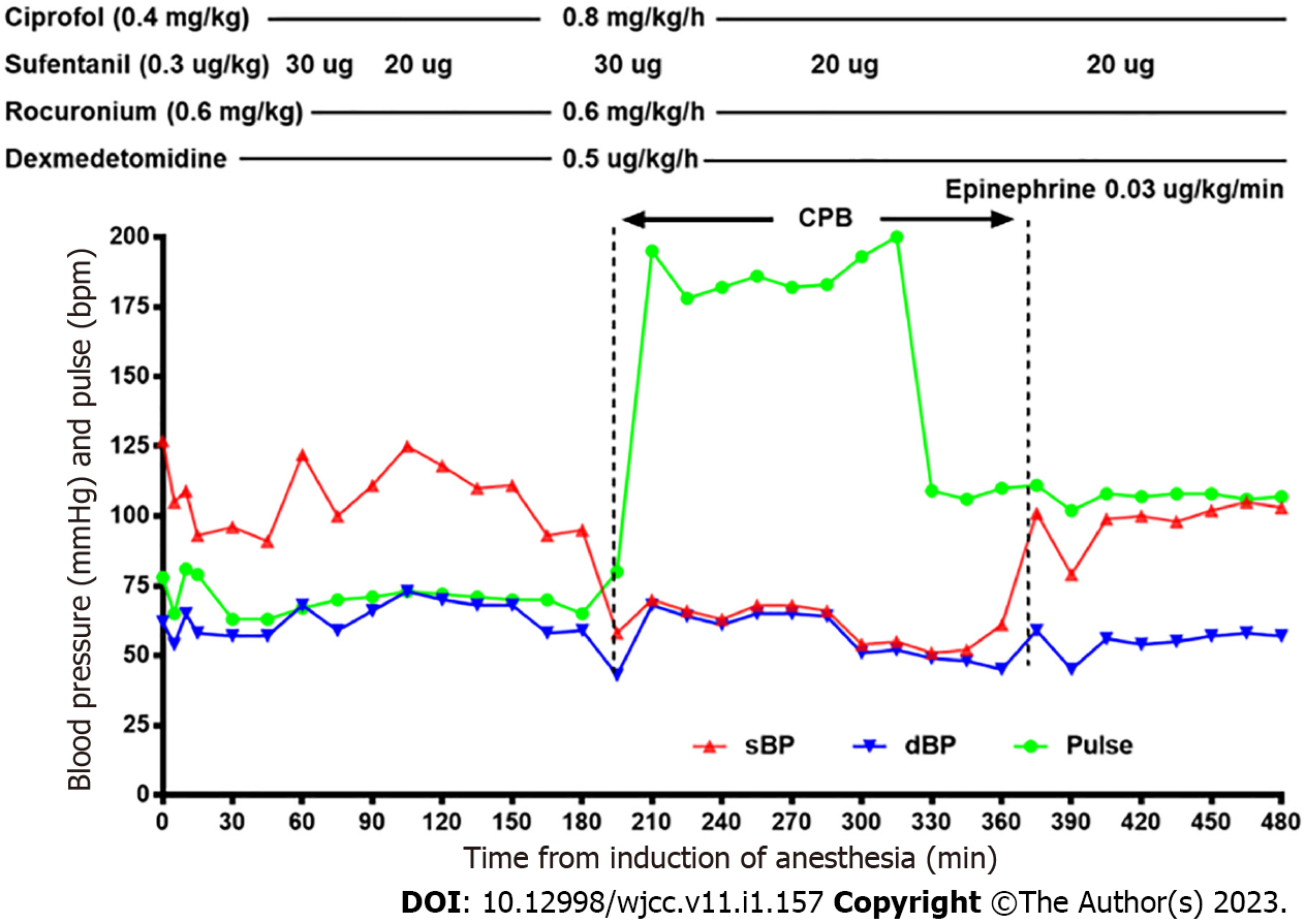
Figure 3 Anesthetic records.
For induction of anesthesia, 0.4 mg/kg ciprofol, 0.3 μg/kg sufentanil, and 0.6 mg/kg rocuronium bromide were administered. Anesthesia was maintained with ciprofol 0.8 mg/kg/h, dexmedetomidine 0.5 μg/kg/h, rocuronium 0.6 mg/kg/h, and intermittent sufentanil. After aortic patency was achieved, 0.03 μg/kg/min epinephrine was used to promote cardiac restart. The total anesthesia time was 480 min, and cardiopulmonary bypass time was 175 min.
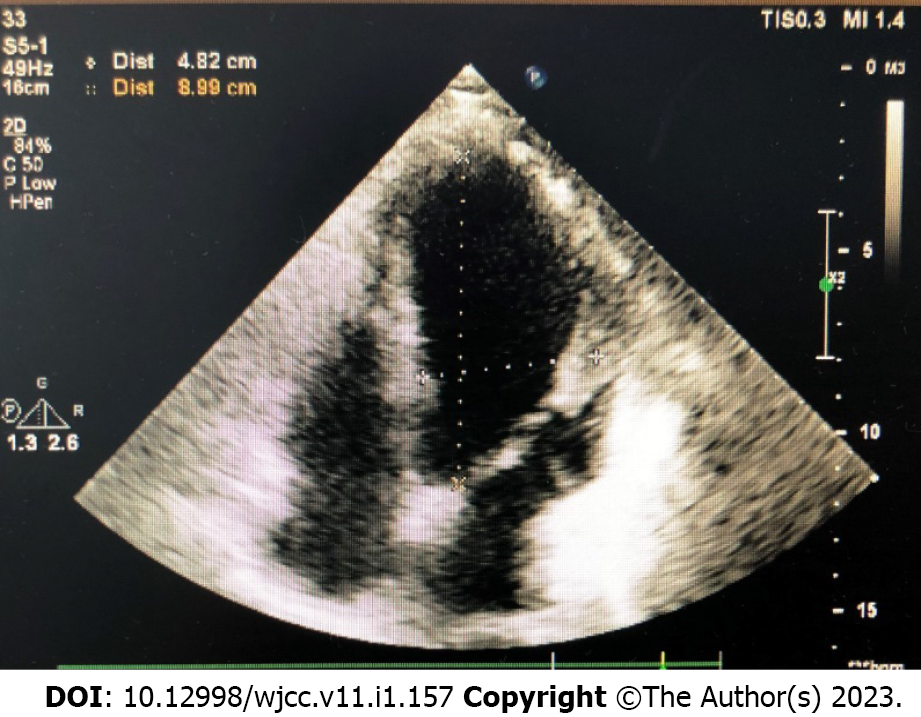
Figure 4 Postoperative echocardiographic images.
The left ventricular volume returned to normal, and the huge apical aneurysm disappeared.
- Citation: Yu L, Bischof E, Lu HH. Anesthesia with ciprofol in cardiac surgery with cardiopulmonary bypass: A case report. World J Clin Cases 2023; 11(1): 157-163
- URL: https://www.wjgnet.com/2307-8960/full/v11/i1/157.htm
- DOI: https://dx.doi.org/10.12998/wjcc.v11.i1.157


