Copyright
©The Author(s) 2023.
World J Clin Cases. Jan 6, 2023; 11(1): 143-149
Published online Jan 6, 2023. doi: 10.12998/wjcc.v11.i1.143
Published online Jan 6, 2023. doi: 10.12998/wjcc.v11.i1.143
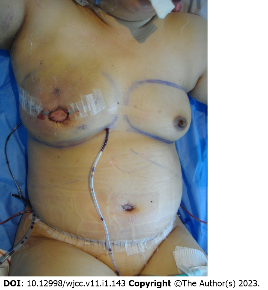
Figure 1 Immediate postoperative view of the deep inferior epigastric artery perforator free flap after total mastectomy of the right breast.
The operation was successful without any events.
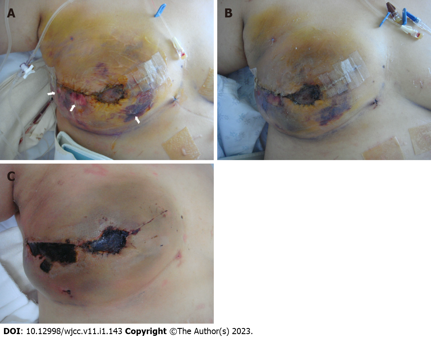
Figure 2 Suffering a deep second to third burn injury.
A: At postoperative day 7, she suffered a deep second to third-degree contact burn with bullae formation (arrows) on the reconstructed right breast due to the incorrect application of a heating pad; B: The burn wound was treated initially with a conventional dressing; C: However, the necrotic changes progressed with eschar formation.
Figure 3 Treatment for burn injury.
A: During eschar resection at postoperative day 21, we could confirm the flap survival by observing the glistening yellow adipose tissue in the subdermal layer and the strongly traceable Doppler sound of the flap pedicle; B: Although we applied a V.A.C. dressing (KCI international, San Antonio, TX.) on the reconstructed right breast to promote burn wound healing, the burn wound worsened, and the exposed adipose tissue changed to dry and brown-colored necrotic tissue after four days of V.A.C. application; C: With further debridement, we observed an obstructed vascular flap pedicle with multiple thrombi in the transferred deep inferior epigastric artery perforator flap tissue; D: After thorough debridement, a bilateral advancement flap was performed and the patient was discharged without any other surgical complications on the 3rd postoperative day.
- Citation: Lim S, Lee DY, Kim B, Yoon JS, Han YS, Eo S. Devastating complication of negative pressure wound therapy after deep inferior epigastric perforator free flap surgery: A case report. World J Clin Cases 2023; 11(1): 143-149
- URL: https://www.wjgnet.com/2307-8960/full/v11/i1/143.htm
- DOI: https://dx.doi.org/10.12998/wjcc.v11.i1.143


