Copyright
©The Author(s) 2022.
World J Clin Cases. Mar 26, 2022; 10(9): 2883-2894
Published online Mar 26, 2022. doi: 10.12998/wjcc.v10.i9.2883
Published online Mar 26, 2022. doi: 10.12998/wjcc.v10.i9.2883
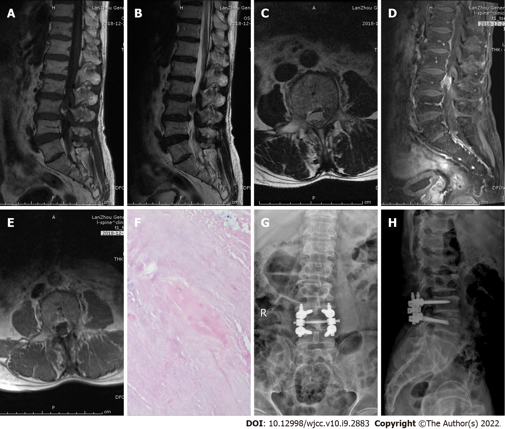
Figure 1 Imaging examination of case 1.
A: T1-weighted preoperative MRI image showing high signal intensity; B and C: Preoperative T2-weighted image showing low signal intensity, and an axial T2-weighted image demonstrating disc fragments in the left posterior epidural space; D and E: Preoperative contrast-enhanced MRI suggested heterogeneous enhancement; F: Postoperative pathology suggested intervertebral disc tissue; G and H: X-ray on postoperative day 2 indicated intact internal fixation.
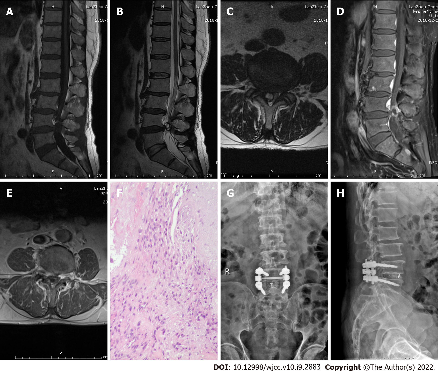
Figure 2 Imaging examination of case 2.
A: T1-weighted preoperative MRI image showing low signal intensity; B and C: Preoperative T2-weighted image showing low signal intensity, and an axial T2-weighted image demonstrating disc fragments in the right posterior epidural space; D and E: Preoperative contrast-enhanced MRI suggested heterogeneous peripheral ring enhancement; F: Postoperative pathology suggested intervertebral disc tissue; G and H: X-ray on postoperative day 2 indicated intact internal fixation.
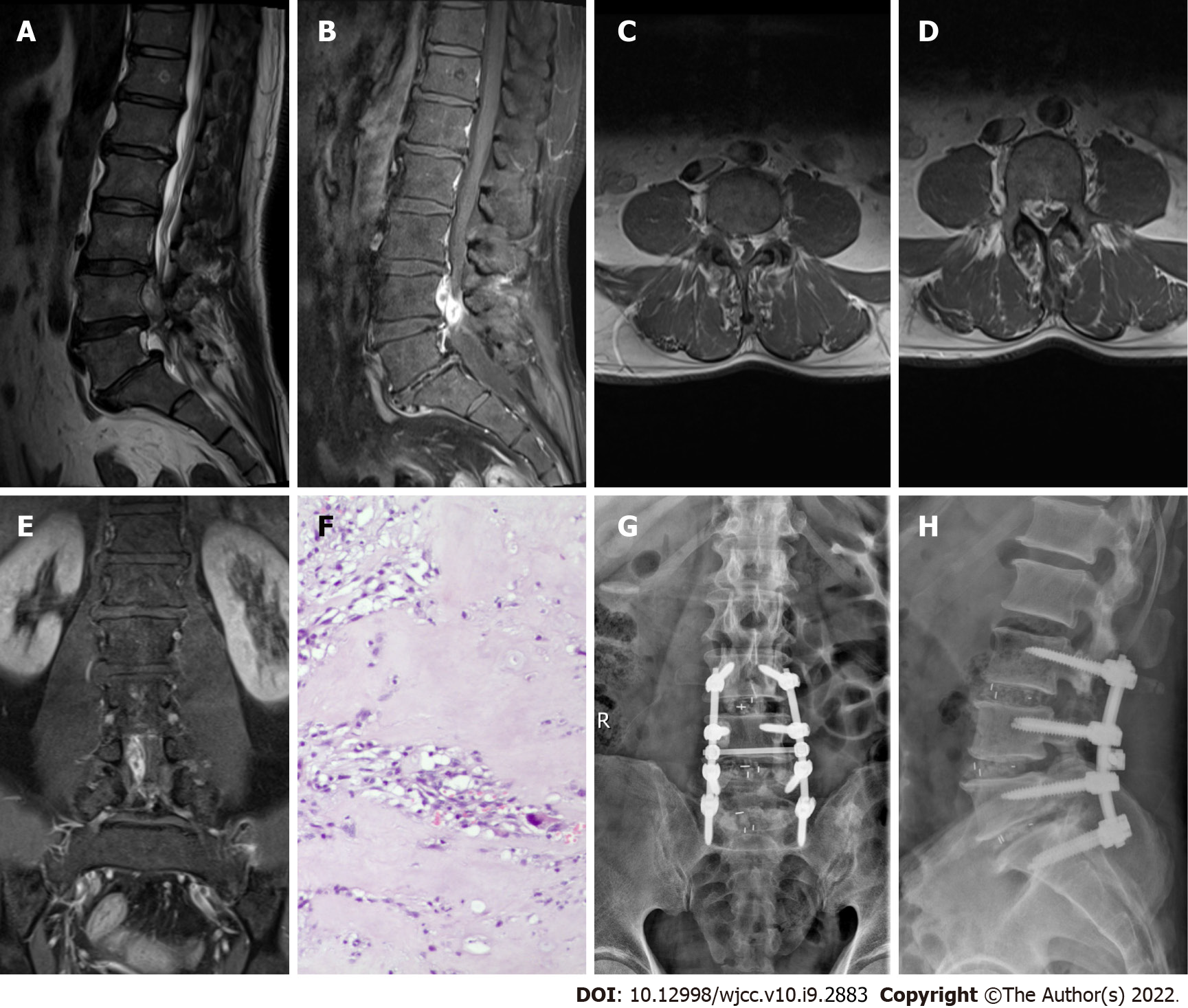
Figure 3 Imaging examination of case 3.
A: T2-weighted preoperative MRI image showing high signal intensity; B–E: Preoperative contrast-enhanced MRI images showing considerable peripheral enhancement, and an axial image demonstrating disc fragments in the right anterior epidural space; F: Postoperative pathology suggested intervertebral disc tissue; G and H: X-ray on postoperative day 2 indicated intact internal fixation.
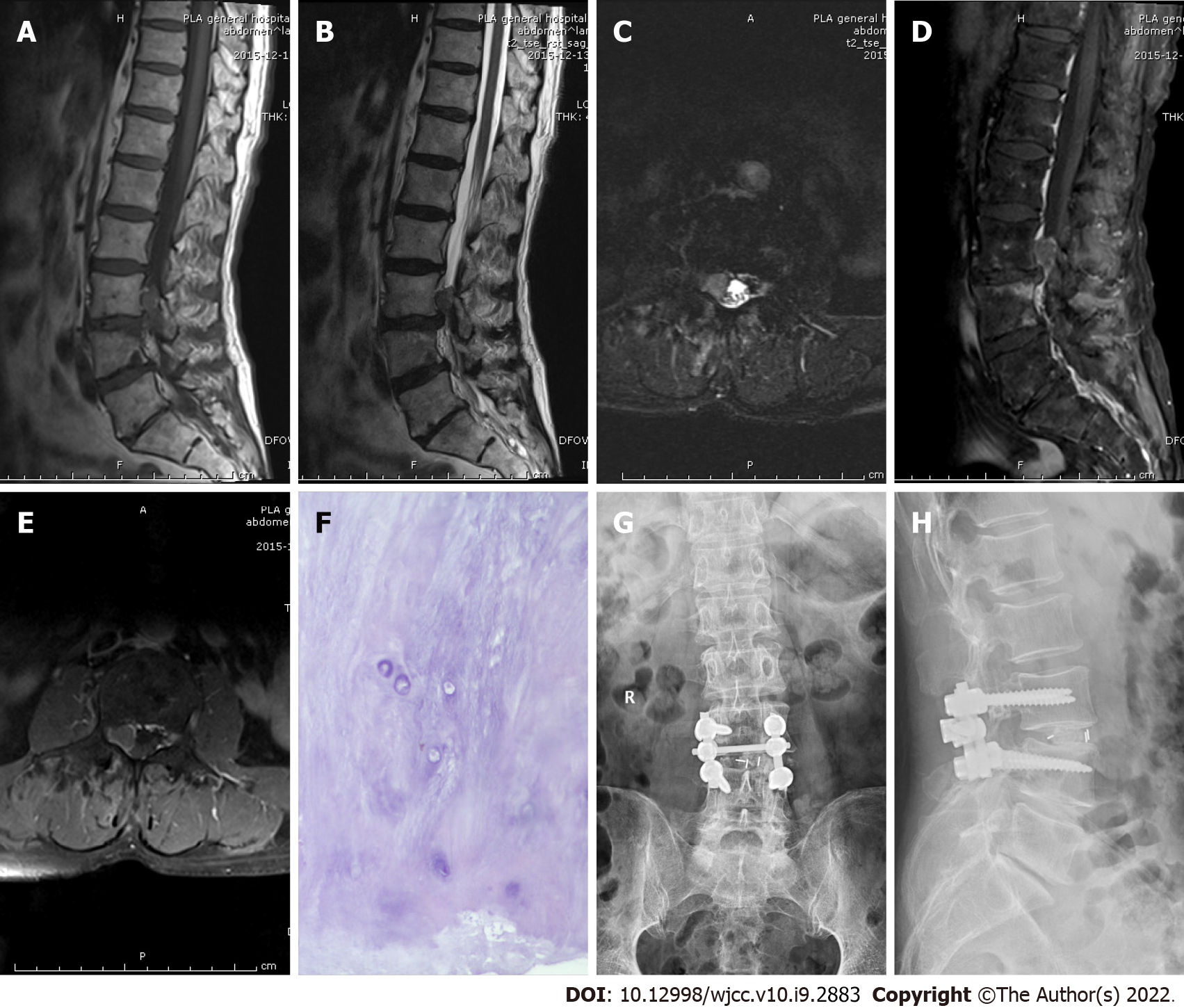
Figure 4 Imaging examination of case 4.
A: Preoperative T1-weighted MRI image showing moderate signal intensity; B and C: Preoperative T2-weighted image showing high signal intensity, and an axial T2-weighted image demonstrating disc fragments in the right anterior epidural space; D and E: Preoperative contrast-enhanced MRI suggested no obvious enhancement; F: Postoperative pathology suggested intervertebral disc tissue; G and H: X-ray on postoperative day 2 indicated intact internal fixation.
- Citation: Li ST, Zhang T, Shi XW, Liu H, Yang CW, Zhen P, Li SK. Lumbar disc sequestration mimicking a tumor: Report of four cases and a literature review. World J Clin Cases 2022; 10(9): 2883-2894
- URL: https://www.wjgnet.com/2307-8960/full/v10/i9/2883.htm
- DOI: https://dx.doi.org/10.12998/wjcc.v10.i9.2883


