Copyright
©The Author(s) 2022.
World J Clin Cases. Mar 26, 2022; 10(9): 2864-2870
Published online Mar 26, 2022. doi: 10.12998/wjcc.v10.i9.2864
Published online Mar 26, 2022. doi: 10.12998/wjcc.v10.i9.2864
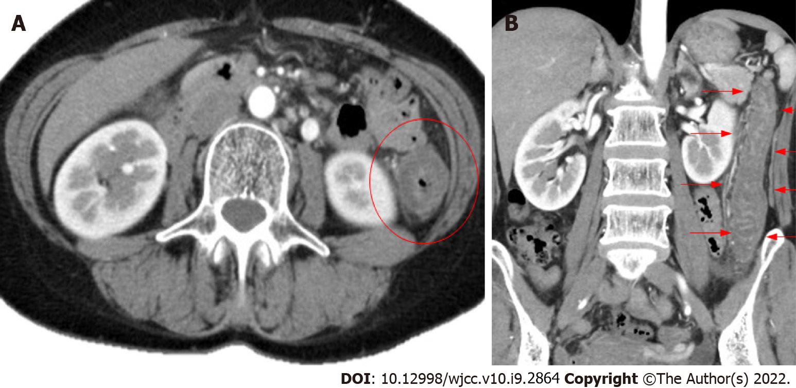
Figure 1 Contrast-enhanced abdominal computed tomography.
A: Horizontal slice; B: Coronal slice edema-like changes are observed from the descending colon to the sigmoid colon (red circle, red arrow).
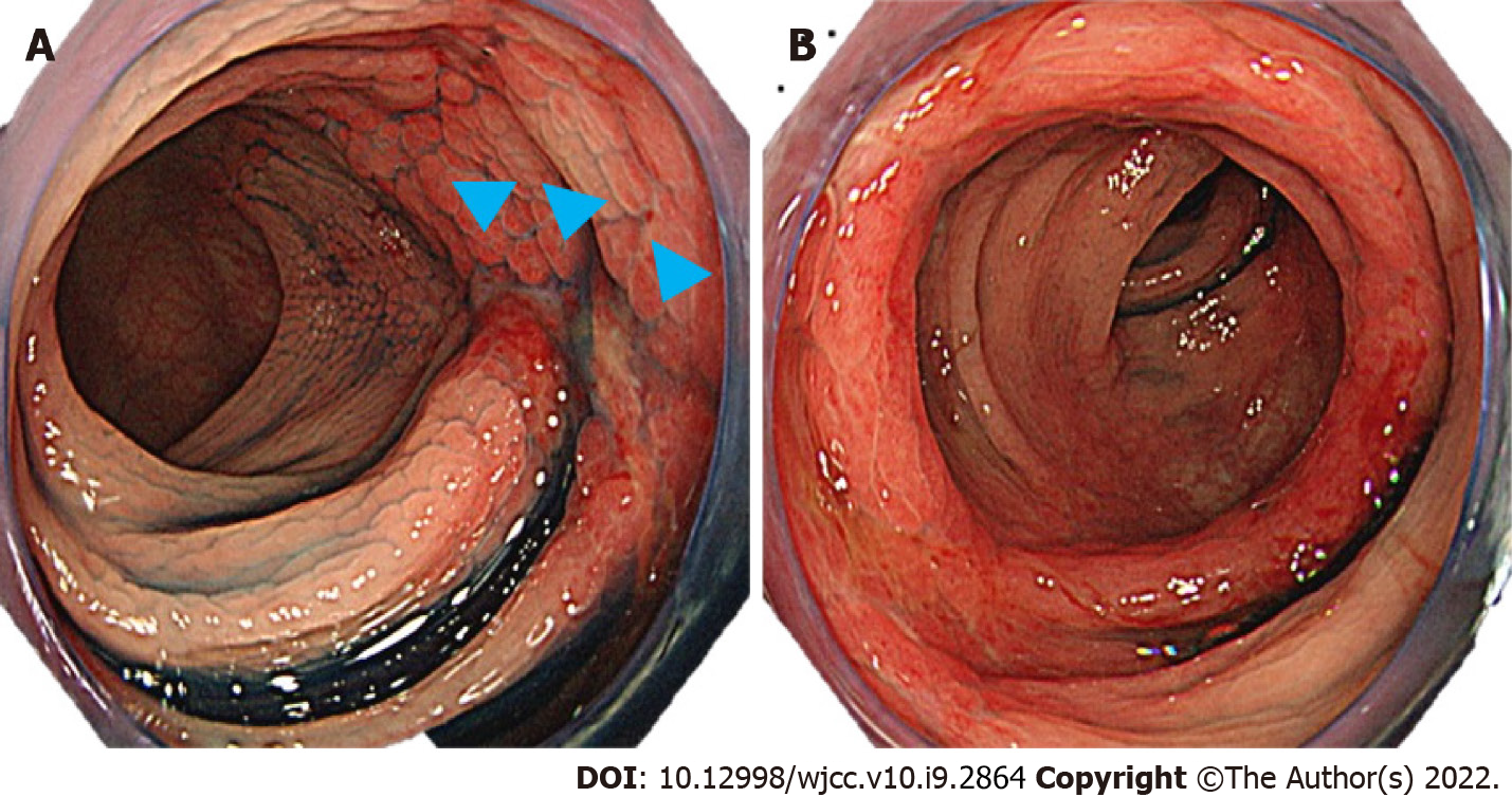
Figure 2 Lower gastrointestinal endoscopy.
A: Longitudinal ulcer (sagittal head); B: Edema-like changes are observed.
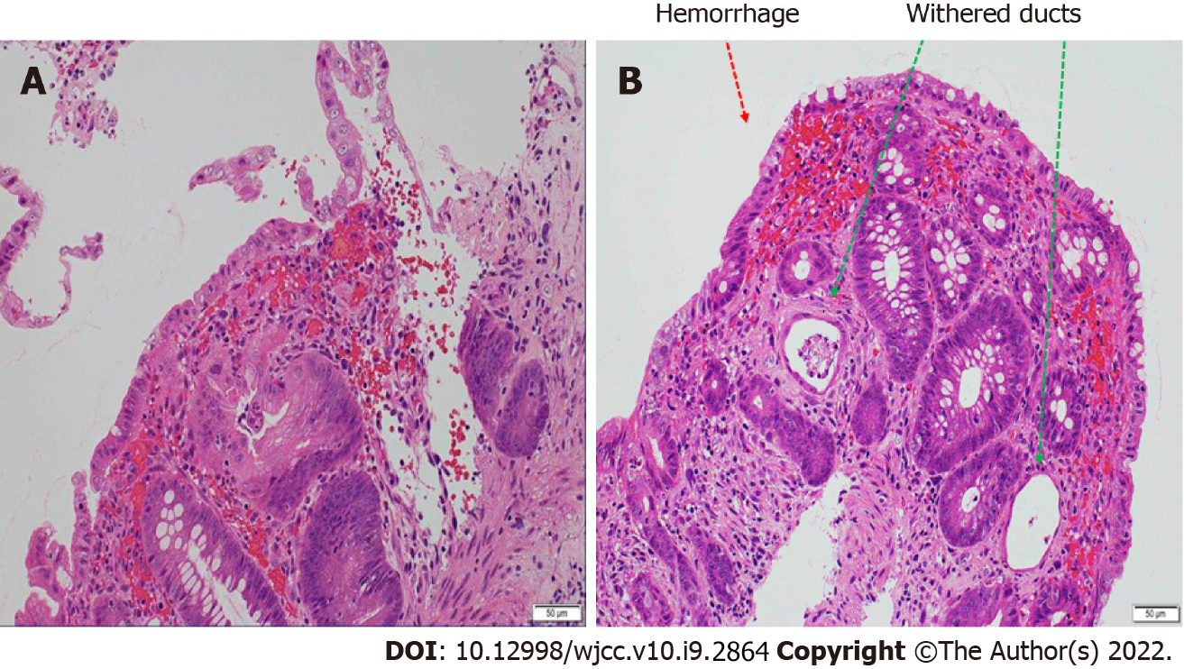
Figure 3 Histopathological examination of lesions in the colon (hematoxylin-eosin staining).
A: Stromal edema and hemorrhage, infiltration of inflammatory cells, and a tendency for detachment of superficial epithelial are observed; B: Withered ducts (green arrow) and hemorrhage (red arrow) are observed.
- Citation: Suzuki C, Kenzaka T. Laninamivir-induced ischemic enterocolitis: A case report. World J Clin Cases 2022; 10(9): 2864-2870
- URL: https://www.wjgnet.com/2307-8960/full/v10/i9/2864.htm
- DOI: https://dx.doi.org/10.12998/wjcc.v10.i9.2864


