Copyright
©The Author(s) 2022.
World J Clin Cases. Mar 26, 2022; 10(9): 2851-2857
Published online Mar 26, 2022. doi: 10.12998/wjcc.v10.i9.2851
Published online Mar 26, 2022. doi: 10.12998/wjcc.v10.i9.2851
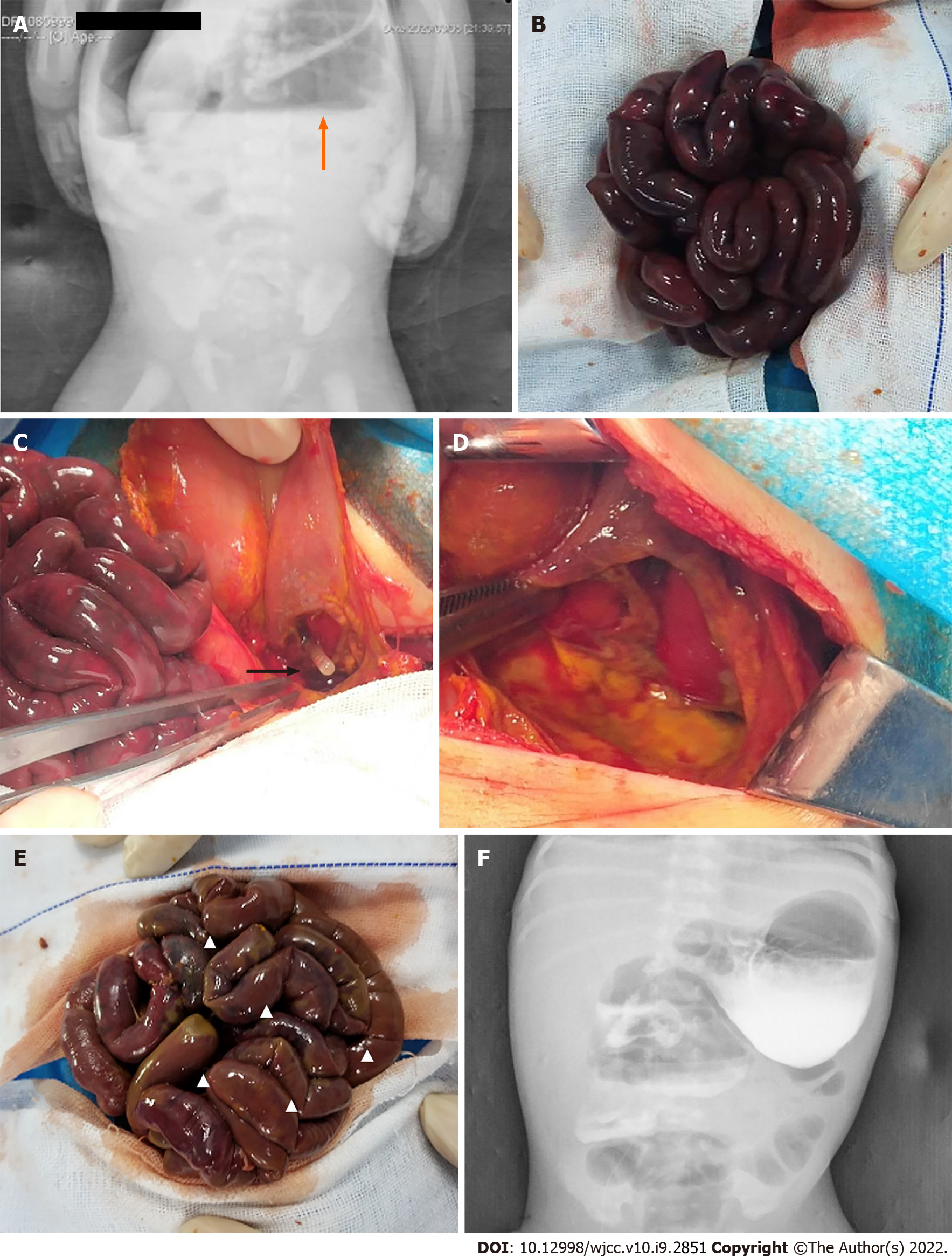
Figure 1 Image examination.
A: Free gases and liquid under the diaphragm; B: The whole bowel loops looked black purplish; C: Defects and perforation of gastric musculature; D: The muscle layer was interrupted at the junction of the normal stomach wall; E: Part of the intestinal canal was left with serosa layer only; F: High density contrast media shadow could be seen in the stomach and part of the intestinal tract.
- Citation: Wang Y, Gu Y, Ma D, Guo WX, Zhang YF. Congenital intestinal malrotation with gastric wall defects causing extensive gut necrosis and short gut syndrome: A case report. World J Clin Cases 2022; 10(9): 2851-2857
- URL: https://www.wjgnet.com/2307-8960/full/v10/i9/2851.htm
- DOI: https://dx.doi.org/10.12998/wjcc.v10.i9.2851


