Copyright
©The Author(s) 2022.
World J Clin Cases. Mar 6, 2022; 10(7): 2351-2356
Published online Mar 6, 2022. doi: 10.12998/wjcc.v10.i7.2351
Published online Mar 6, 2022. doi: 10.12998/wjcc.v10.i7.2351
Figure 1 Diffusion-weighted imaging.
Case 1, A and B: Diffusion-weighted imaging and perfusion-weighted imagingshow acute infarctions in the left temporal and insular lobes with no significant diffusion-weighted imaging-perfusion-weighted imaging mismatch, C: Magnetic resonance angiography shows occlusion of the M2 inferior trunk. Case 2, D and E: Diffusion-weighted imaging and perfusion-weighted imaging show acute infarctions in the left parietal and insular lobes with significant DWI-PWI mismatch; F: Digital subtraction angiography confirmed the occlusion of the M2 inferior trunk; G: After thrombectomy, reperfusion and good antegrade blood flow was confirmed.
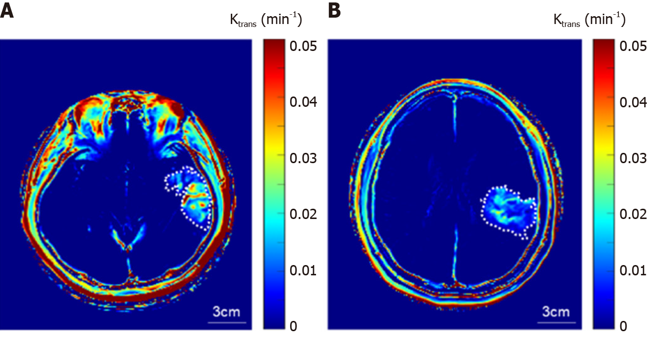
Figure 2 Dynamic contrast-enhanced magnetic resonance imaging.
A representative blood-brain barrier permeability (Ktrans) map of the patients without (A) and with (B) thrombectomy. The dashed line indicates the manually segmented stroke region with enhanced Ktrans.
- Citation: Seo Y, Kim J, Chang MC, Huh H, Lee EH. Relationship between treatment types and blood–brain barrier disruption in patients with acute ischemic stroke: Two case reports. World J Clin Cases 2022; 10(7): 2351-2356
- URL: https://www.wjgnet.com/2307-8960/full/v10/i7/2351.htm
- DOI: https://dx.doi.org/10.12998/wjcc.v10.i7.2351


