Copyright
©The Author(s) 2022.
World J Clin Cases. Mar 6, 2022; 10(7): 2341-2350
Published online Mar 6, 2022. doi: 10.12998/wjcc.v10.i7.2341
Published online Mar 6, 2022. doi: 10.12998/wjcc.v10.i7.2341
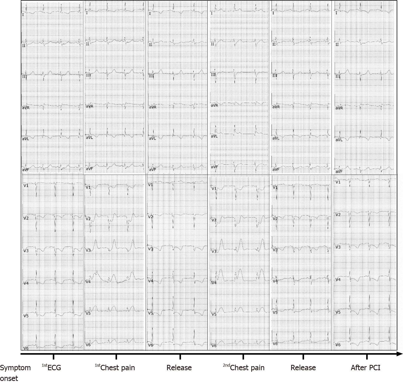
Figure 1 The dynamic changes in the electrocardiogram of the patient from symptom onset to discharge from the hospital.
ECG: Electrocardiogram; PCI: Percutaneous coronary intervention.
Figure 2 Percutaneous coronary intervention for occlusion of the left main coronary artery.
A: Left coronary angiography using a pigtail catheter placed in the left coronary sinus revealed occlusion of the proximal left main trunk coronary artery (LMT); B: An everolimus-eluting stent (3.5 mm × 24 mm) was implanted into the focal lesions of the LMT; C: LMT angiography after implantation of an everolimus-eluting stent (Boston Scientific, MN, United States).
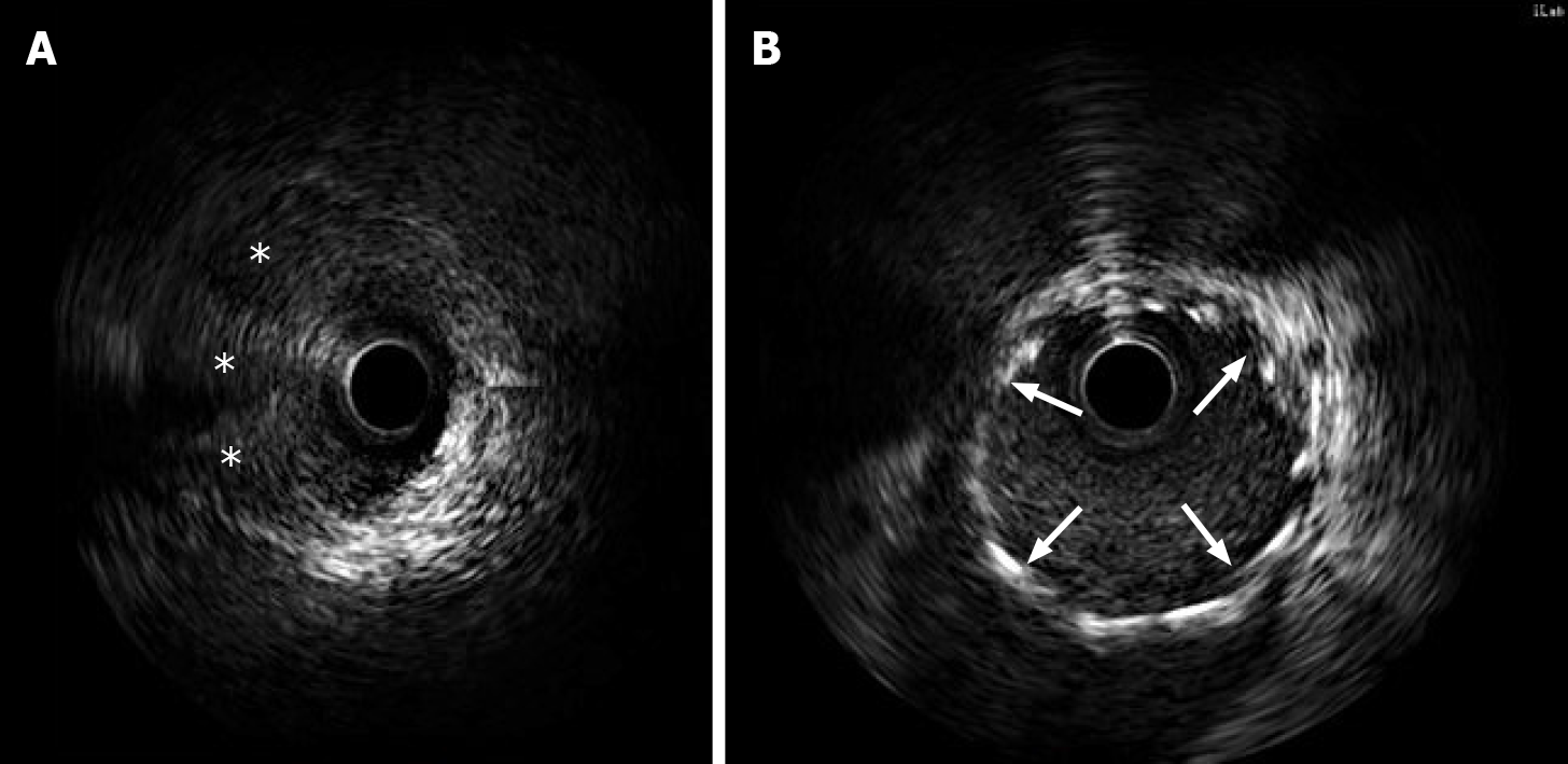
Figure 3 Intravascular ultrasound confirmed a false lumen with a prominent dissection from the left main trunk coronary artery to the ostium of the left anterior descending artery.
A: Intravascular ultrasound (IVUS) images showed artery dissection starting from the left main trunk coronary artery to the ostium of the left anterior descending artery. The guidewire passed through the true lumen properly; B: IVUS demonstrated stents attached to the endothelium well that covered the artery dissections completely. Intramural hematoma (stellate). The implanted stents are indicated with arrows.
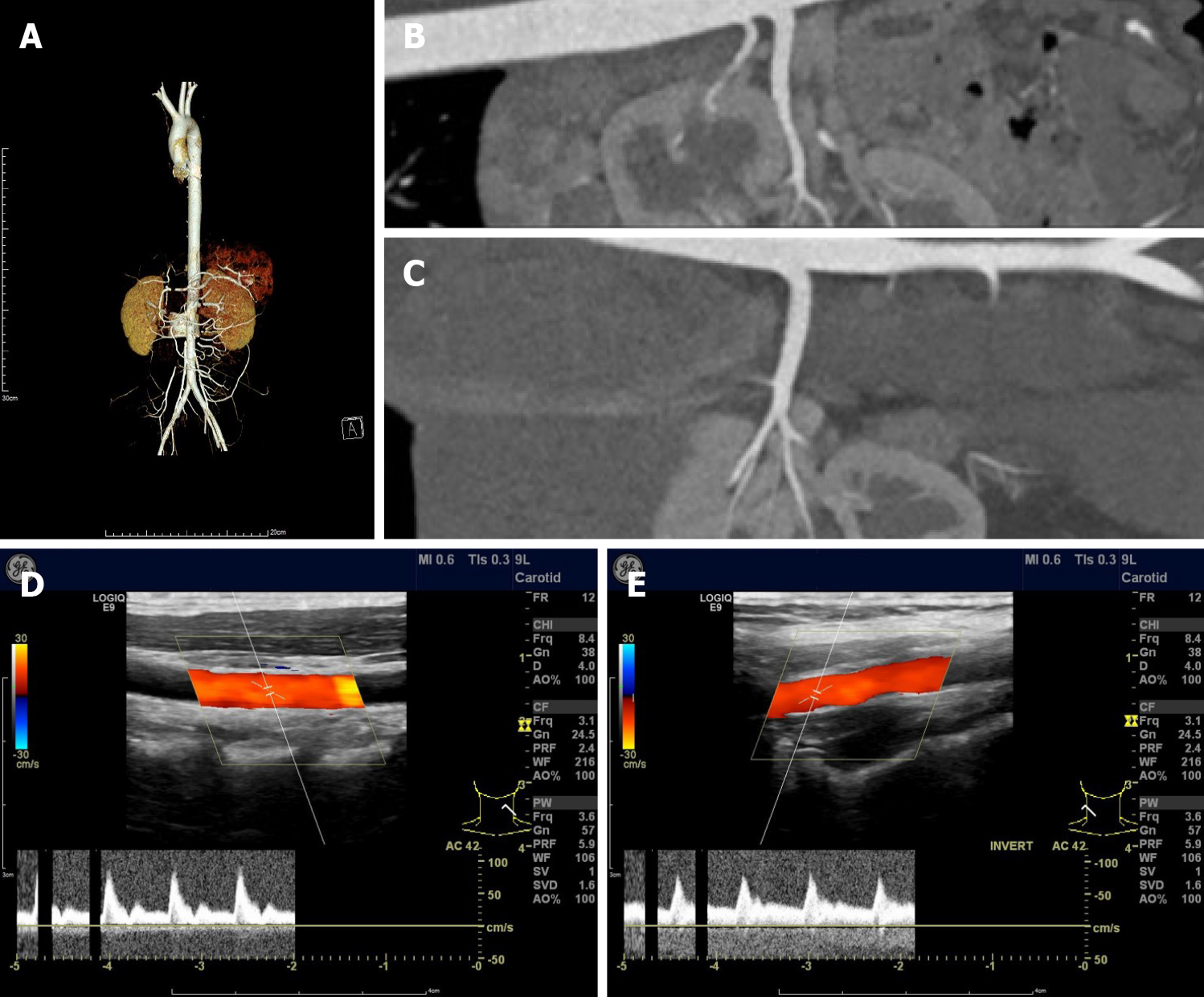
Figure 4 Arteriogram with contrast and vascular ultrasonography showed no sign of fibromuscular dysplasia.
A: Arteriogram with contrast of the aorta; B: Arteriogram with contrast of the middle right; C: Arteriogram with contrast of left renal arteries; D: Vascular ultrasonography of the left; E: Vascular ultrasonography of right carotid arteries.
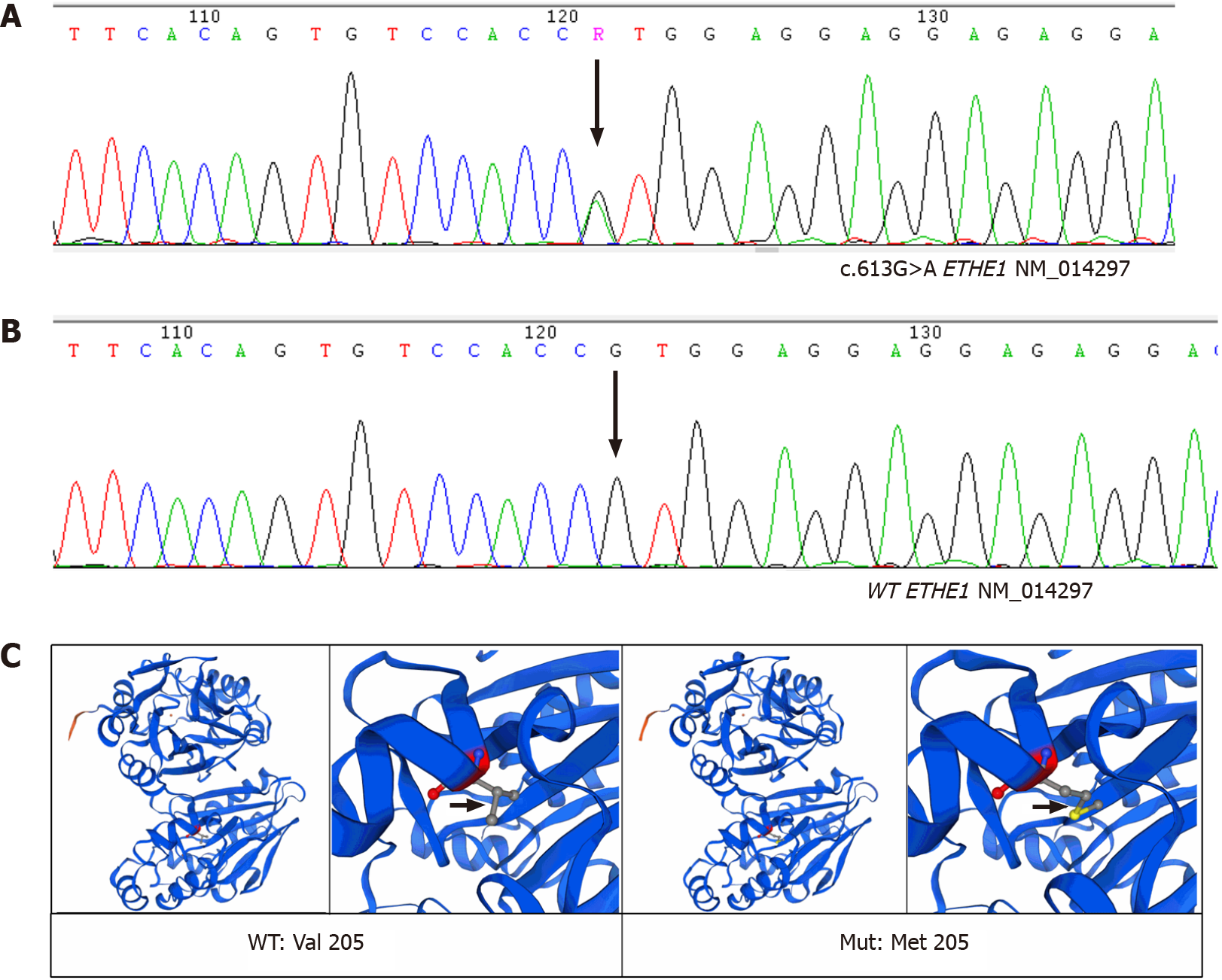
Figure 5 A novel heterozygous missense variant in the ETHE1 gene was found.
A: Sanger sequencing confirmed that the patient was heterozygous for the variant ETHE1 c.613G>A (p.Val205Met), the arrow indicated the position of the variant; B: Sanger sequencing confirmed that no ETHE1 variant was found in her parents or her younger sister. C: Structural models of wild-type and mutant ETHE1 were built using SWISSMODEL. The position of the variant was indicated by arrows.
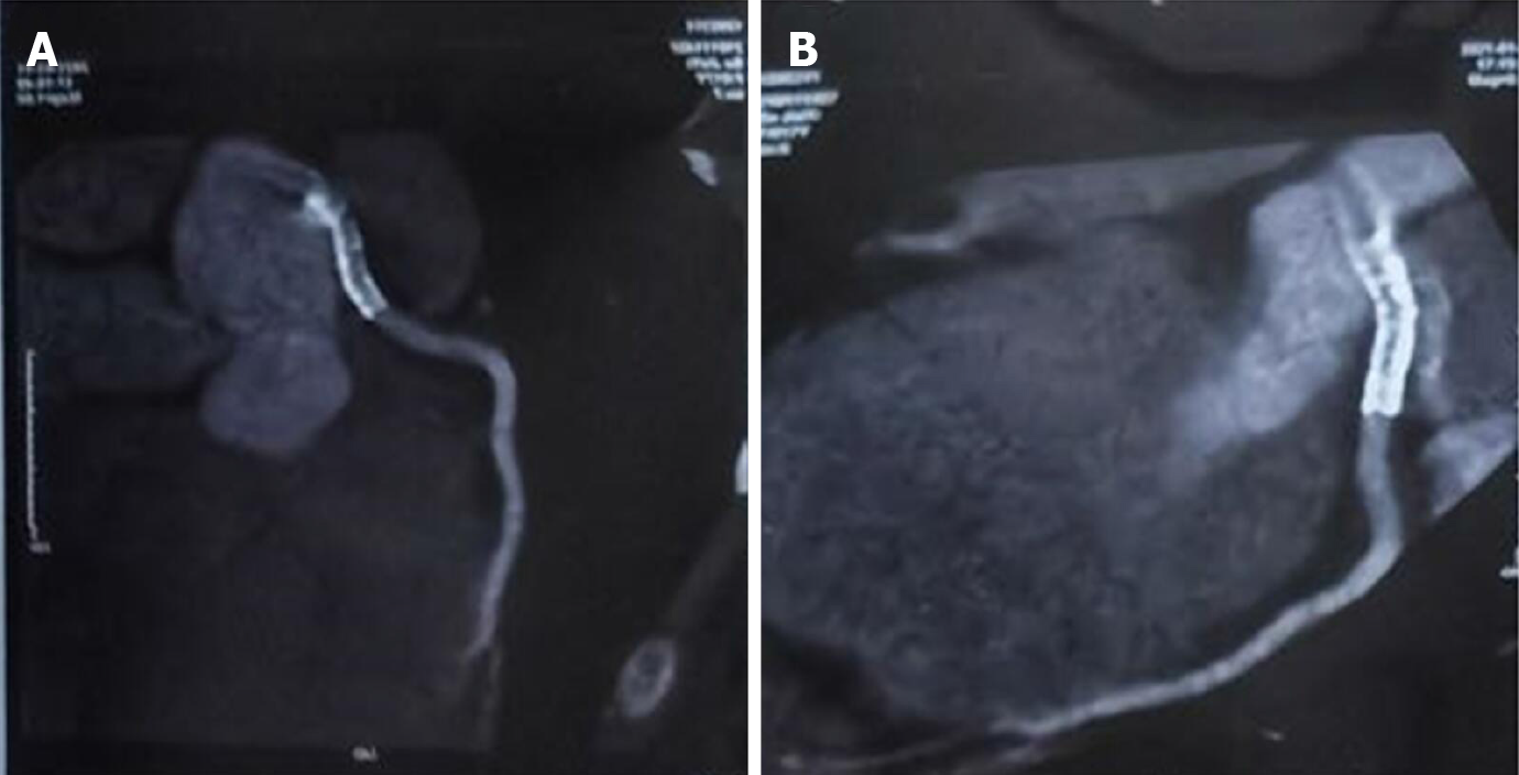
Figure 6
Coronary computed tomography at 12 month.
A and B: Follow-up showed no in-stent restenosis.
Figure 7 Case report timeline.
PCI: Percutaneous coronary intervention; IABP: Intra-aortic balloon pump; IVUS: Intravascular ultrasonography.
- Citation: Liu SF, Zhao YN, Jia CW, Ma TY, Cai SD, Gao F. Spontaneous dissection of proximal left main coronary artery in a healthy adolescent presenting with syncope: A case report. World J Clin Cases 2022; 10(7): 2341-2350
- URL: https://www.wjgnet.com/2307-8960/full/v10/i7/2341.htm
- DOI: https://dx.doi.org/10.12998/wjcc.v10.i7.2341


