Copyright
©The Author(s) 2022.
World J Clin Cases. Mar 6, 2022; 10(7): 2307-2314
Published online Mar 6, 2022. doi: 10.12998/wjcc.v10.i7.2307
Published online Mar 6, 2022. doi: 10.12998/wjcc.v10.i7.2307
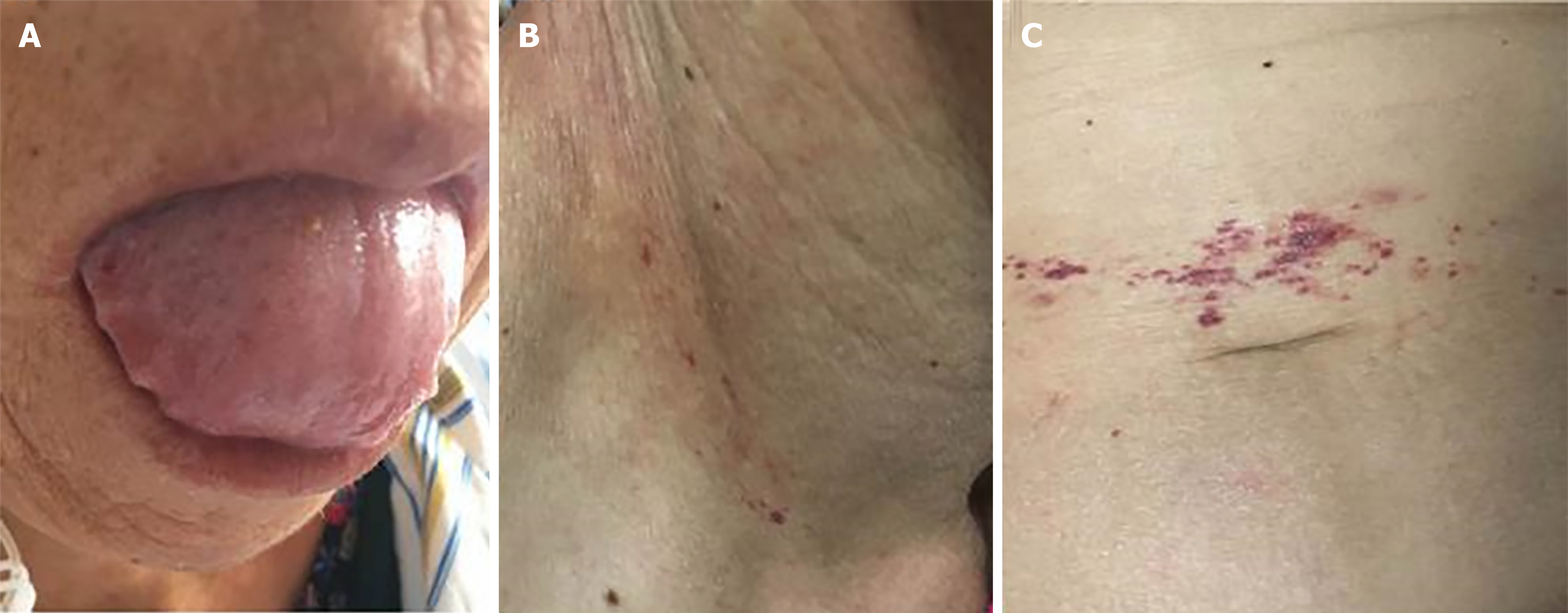
Figure 1 Physical examination.
A: Swollen tongue with teeth prints; B: Skin purpura over the right neck; C: Skin purpura around the umbilicus.
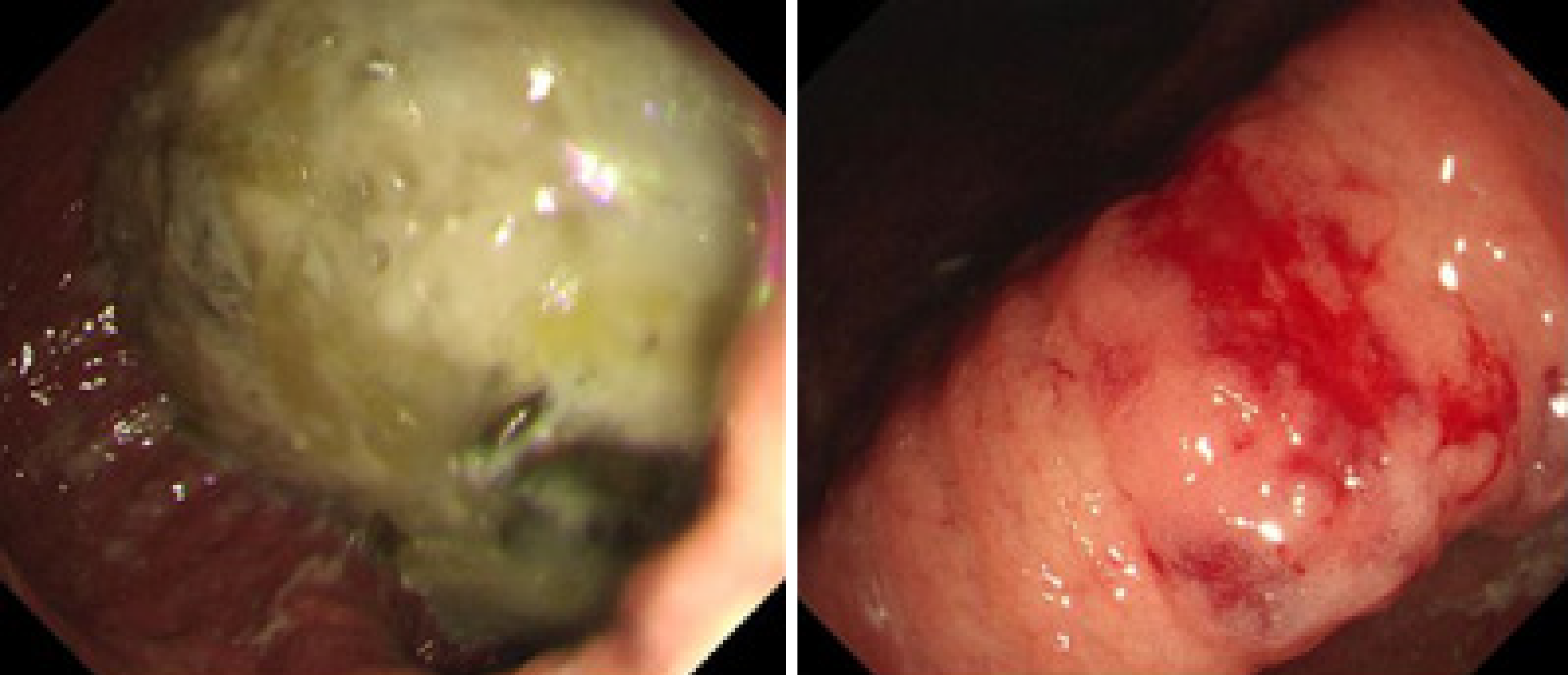
Figure 2
Gastroscopy showing gastric retention, gastric angular mucosal coarseness, hyperemia, and mild oozing of blood.
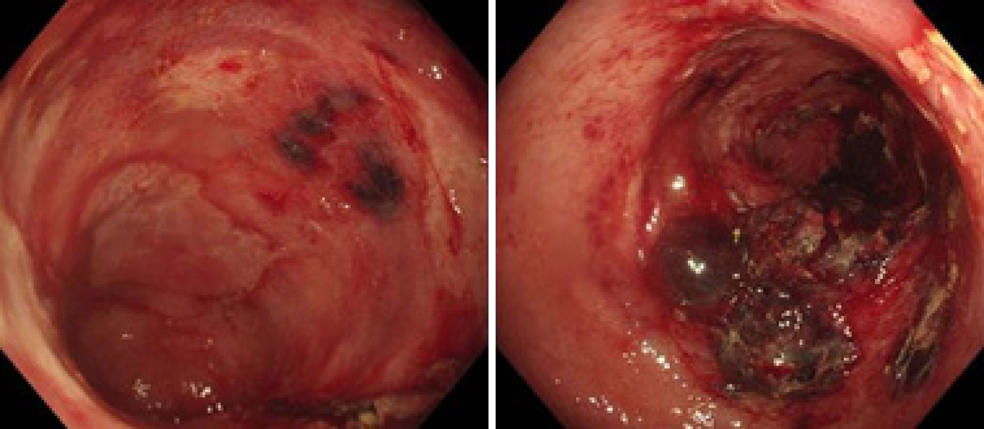
Figure 3
Colonoscopy showing mucosal hyperemia, edema with multiple ovals, irregular ulcers, ecchymosis, and hematoma involving the descending colon and sigmoid colon.
Figure 4
Congo red staining revealed positively staining deposits in the lamina propria of the colon without plasmacytic infiltration (× 400).
- Citation: Liu AL, Ding XL, Liu H, Zhao WJ, Jing X, Zhou X, Mao T, Tian ZB, Wu J. Gastrointestinal amyloidosis in a patient with smoldering multiple myeloma: A case report . World J Clin Cases 2022; 10(7): 2307-2314
- URL: https://www.wjgnet.com/2307-8960/full/v10/i7/2307.htm
- DOI: https://dx.doi.org/10.12998/wjcc.v10.i7.2307


