Copyright
©The Author(s) 2022.
World J Clin Cases. Mar 6, 2022; 10(7): 2301-2306
Published online Mar 6, 2022. doi: 10.12998/wjcc.v10.i7.2301
Published online Mar 6, 2022. doi: 10.12998/wjcc.v10.i7.2301
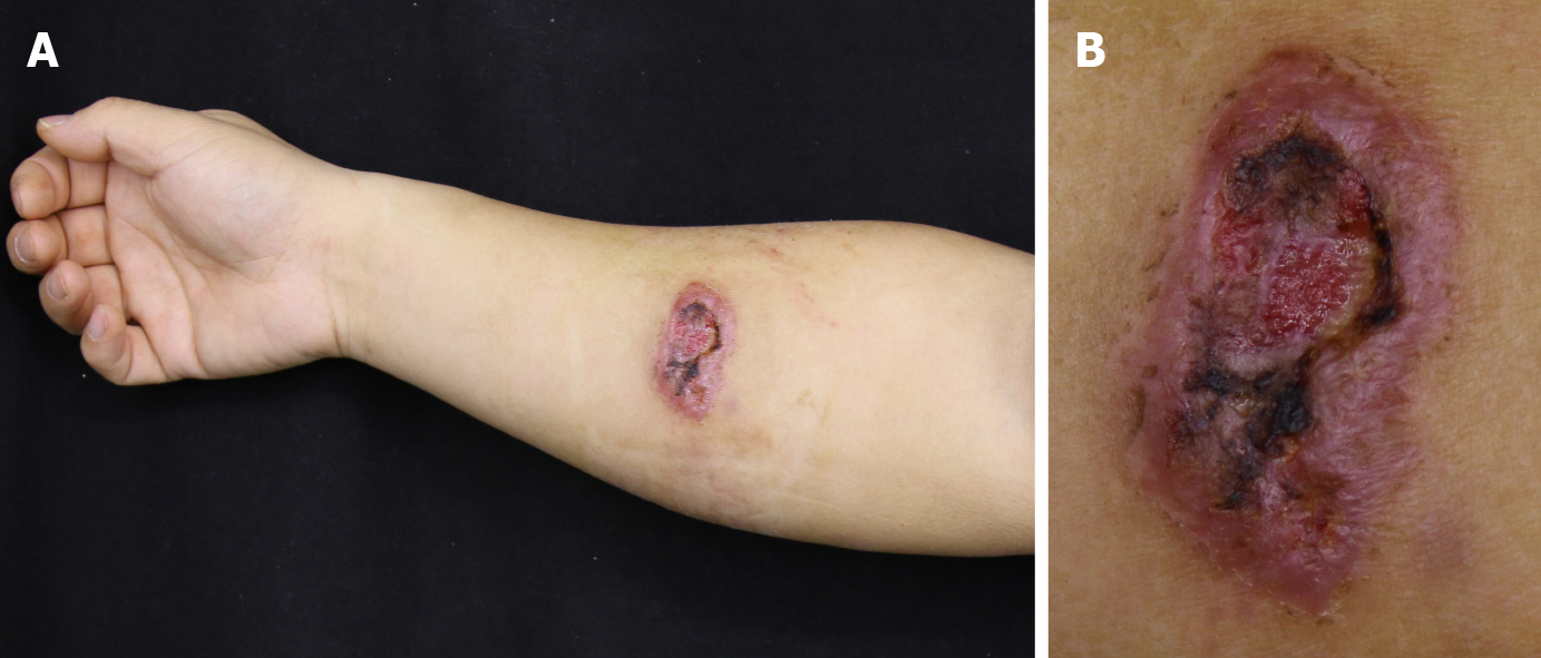
Figure 1 Picture of the lesion.
A: Overall view of the skin plaque with rupture on the right forearm; B: Local magnification of the right forearm plaque with ulcer. The black crust and translucent fluid were visible.
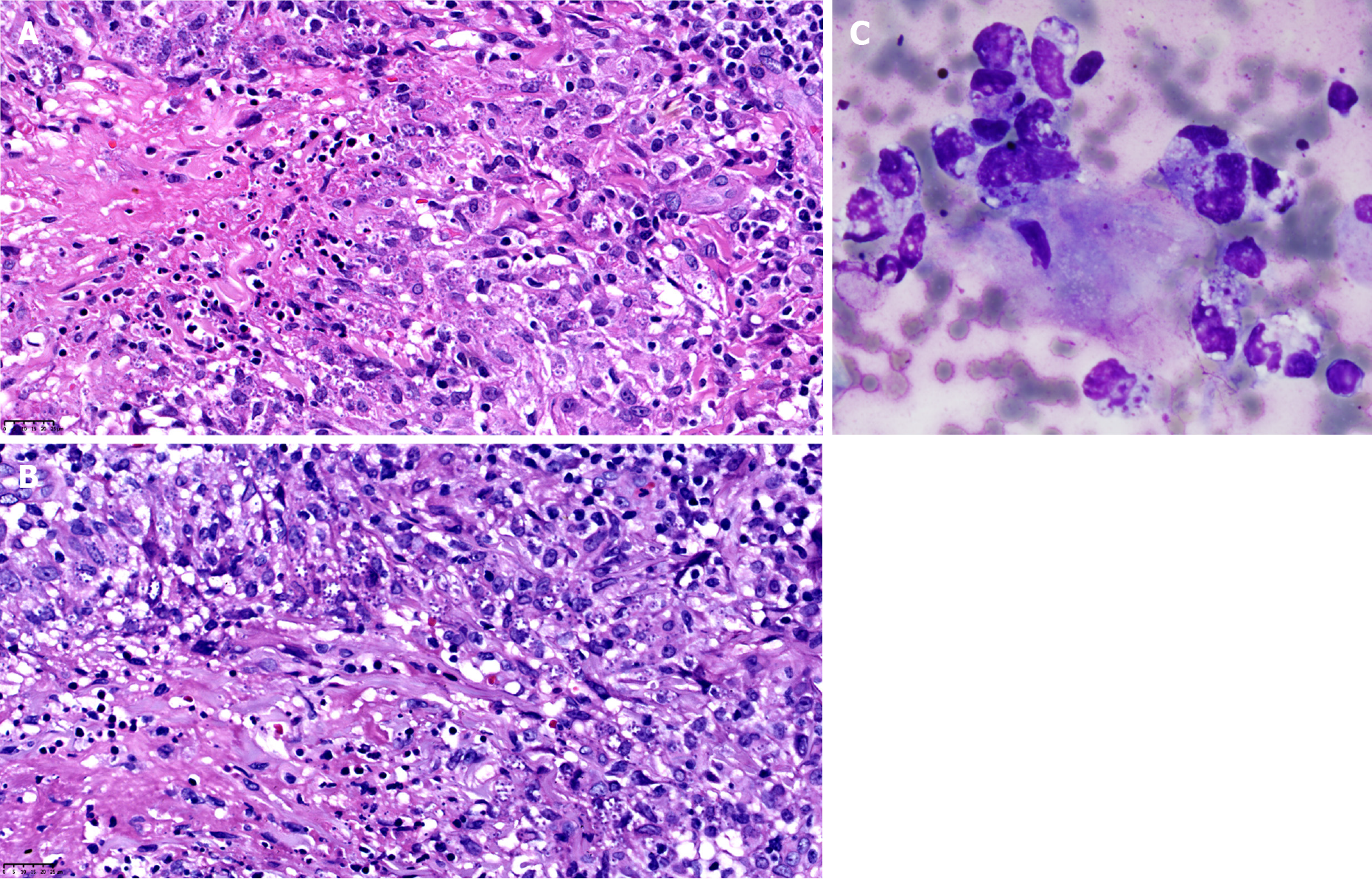
Figure 2 Pathological examination of the lesion.
A: Hematoxylin and eosin section shows a large number of granular structures within the epithelial cells (40 ×); B: Giemsa staining revealed blue flagellar free bodies in tissue cells (40 ×); C: Giemsa staining revealed blue flagellar-free bodies in tissue cells of the secretion smear (100 ×).
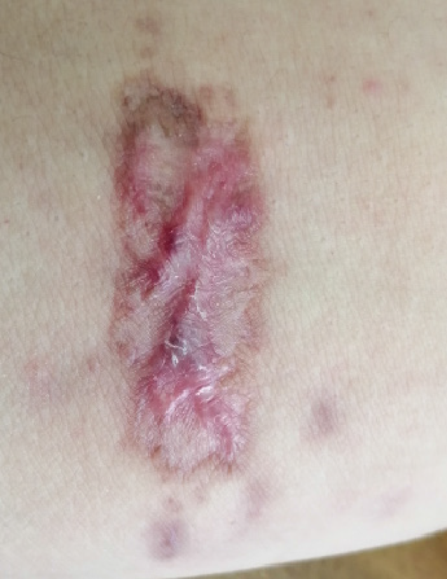
Figure 3 Post-treatment follow-up.
The plaques disappeared, the ulcer healed, and a scar remained.
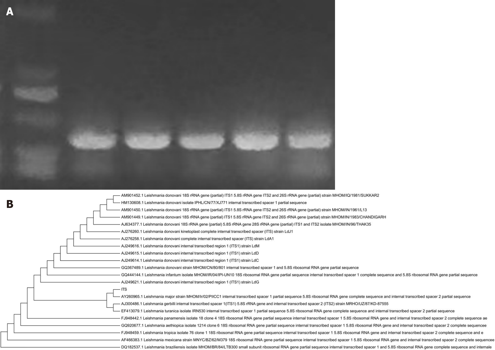
Figure 4 Pathogen identification test.
A: Polymerase chain reaction (PCR) amplification of ITS1. The PCR product can be seen as a single band with a size of 336 bp; B: Phylogenetic tree, in which ITS fetching sequences were the closest relatives to Leishmania major.
- Citation: Zhuang L, Su J, Tu P. Cutaneous leishmaniasis presenting with painless ulcer on the right forearm: A case report. World J Clin Cases 2022; 10(7): 2301-2306
- URL: https://www.wjgnet.com/2307-8960/full/v10/i7/2301.htm
- DOI: https://dx.doi.org/10.12998/wjcc.v10.i7.2301


