Copyright
©The Author(s) 2022.
World J Clin Cases. Mar 6, 2022; 10(7): 2275-2280
Published online Mar 6, 2022. doi: 10.12998/wjcc.v10.i7.2275
Published online Mar 6, 2022. doi: 10.12998/wjcc.v10.i7.2275
Figure 1 Preoperative imaging examination.
A: Preoperative brain computed tomography revealed significantly dilated supratentorial ventricles, capped edema at bilateral frontal angles, huge occupying cysts in the right cerebellar hemisphere, and compression displacement in the right cerebellar hemisphere and the four ventricles; B and C: Preoperative magnetic resonance imaging T1 and T2 scans revealed significantly dilated lateral ventricle, capped edema at bilateral frontal angles, huge occupying cysts in the right cerebellar hemisphere and compression displacement in the right cerebellar hemisphere and the four ventricles; D: The sagittal position of cranial enhanced magnetic resonance also shows the above signs.
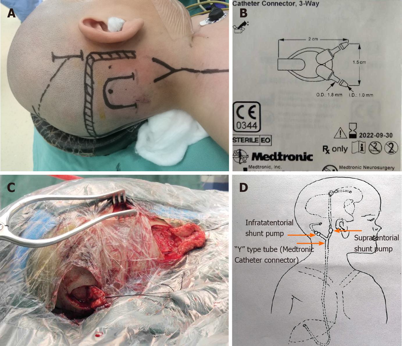
Figure 2 Surgical position, method.
A: Patient position and incision design during surgery; B: Medtronic catheter connector; C: Intraoperative incision; D: A supratentorial adjustable pump was placed behind the ear, connected to a three-way valve; a drainage tube for cerebellar hemispheric cysts was connected to the ordinary non-adjustable pump, and then connected to the three-way valve. The other end of the three-way valve was connected to the end of the abdominal cavity, as in general shunt therapy. Citation for Figure 2B: The authors have obtained the permission for figure using from the Medtronic (see Supplementary material).
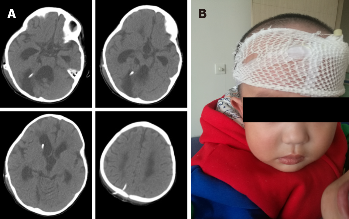
Figure 3 Postoperative imaging examination and child status.
A: Postoperative brain computed tomography showing shrunk lateral ventricles and disappeared capped edema at the bilateral frontal angle; the head ends of the drainage tube in the lateral ventricle and posterior fossa cysts were well-positioned; B: Postoperative status of the child: Brain computed tomography at 1 mo postoperatively revealed significantly shrunk lateral ventricle; the head ends of the drainage tube in the lateral ventricle and posterior fossa cysts were well-positioned. The cysts were significantly shrunk and there was cerebellar vermis dysplasia.
- Citation: Dong ZQ, Jia YF, Gao ZS, Li Q, Niu L, Yang Q, Pan YW, Li Q. Y-shaped shunt for the treatment of Dandy-Walker malformation combined with giant arachnoid cysts: A case report. World J Clin Cases 2022; 10(7): 2275-2280
- URL: https://www.wjgnet.com/2307-8960/full/v10/i7/2275.htm
- DOI: https://dx.doi.org/10.12998/wjcc.v10.i7.2275


