Copyright
©The Author(s) 2022.
World J Clin Cases. Mar 6, 2022; 10(7): 2253-2260
Published online Mar 6, 2022. doi: 10.12998/wjcc.v10.i7.2253
Published online Mar 6, 2022. doi: 10.12998/wjcc.v10.i7.2253
Figure 1 Enhanced computed tomography findings.
A: A huge soft tissue mass was found in the patient’s right upper abdomen; B: A portion of the mass showed an inhomogeneous enhancement in the arterial phase; C: A tissue mass with heterogeneous density was found in the hepatogastric space and no free air was detected.
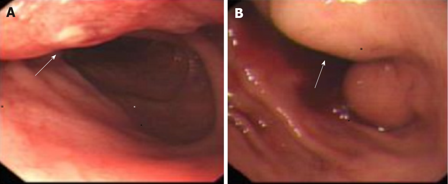
Figure 2 Endoscopic findings.
A: An ulceration was observed in the gastric antrum mucosa; B: Deformation of the gastric sinus and duodenal bulb was visualized.
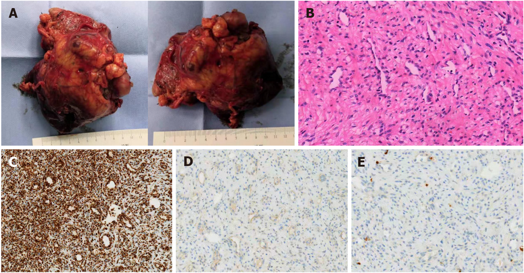
Figure 3 Pathological examinations of the tumor.
A: An 11 cm × 7 cm × 5 cm tumor as seen on the serosa of gastro-duodenal junction, with some necrotic rupture and cystic degeneration visible; B: The tumor was composed of spindle cells with clarified cytoplasm, accompanied by myxoid stroma and arborizing blood vessels (hematoxylin/eosin stain, × 200 magnification); C: Immunohistochemistry staining results were positivity for succinate dehydrogenase subunit B (× 200 magnification); D: Immunohistochemistry staining results were negativity for DOG-1 (× 200 magnification); E: Immunohistochemistry staining results were negativity for CD117 (× 200 magnification).
Figure 4 Images from the enhanced computed tomography review.
A: Following mass removal, there were no signs of tumor recurrence; B: No abnormality as apparent in the arterial phase.
- Citation: Zhang R, Xia LG, Huang KB, Chen ND. Huge gastric plexiform fibromyxoma presenting as pyemia by rupture of tumor: A case report. World J Clin Cases 2022; 10(7): 2253-2260
- URL: https://www.wjgnet.com/2307-8960/full/v10/i7/2253.htm
- DOI: https://dx.doi.org/10.12998/wjcc.v10.i7.2253


