Copyright
©The Author(s) 2022.
World J Clin Cases. Mar 6, 2022; 10(7): 2229-2246
Published online Mar 6, 2022. doi: 10.12998/wjcc.v10.i7.2229
Published online Mar 6, 2022. doi: 10.12998/wjcc.v10.i7.2229
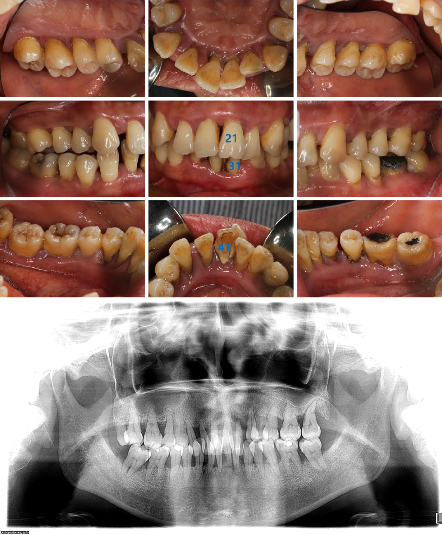
Figure 1 Intraoral images and radiographic image obtained at the first visit.
Intraoral images show the poor level of oral hygiene, a large amount of dental calculus, and obvious plaque retention. No. 21 tooth was distinctly labially inclined and Nos. 31 and 41 were extremely loose. The radiographic image reveals that the full mouth alveolar bone was absorbed to varying degrees into the middle 1/2 and apical 1/3 of the root, with Nos. 22 and 31 absorbed to the apical region.
Figure 2
Timeline flow chart of the treatment plan.
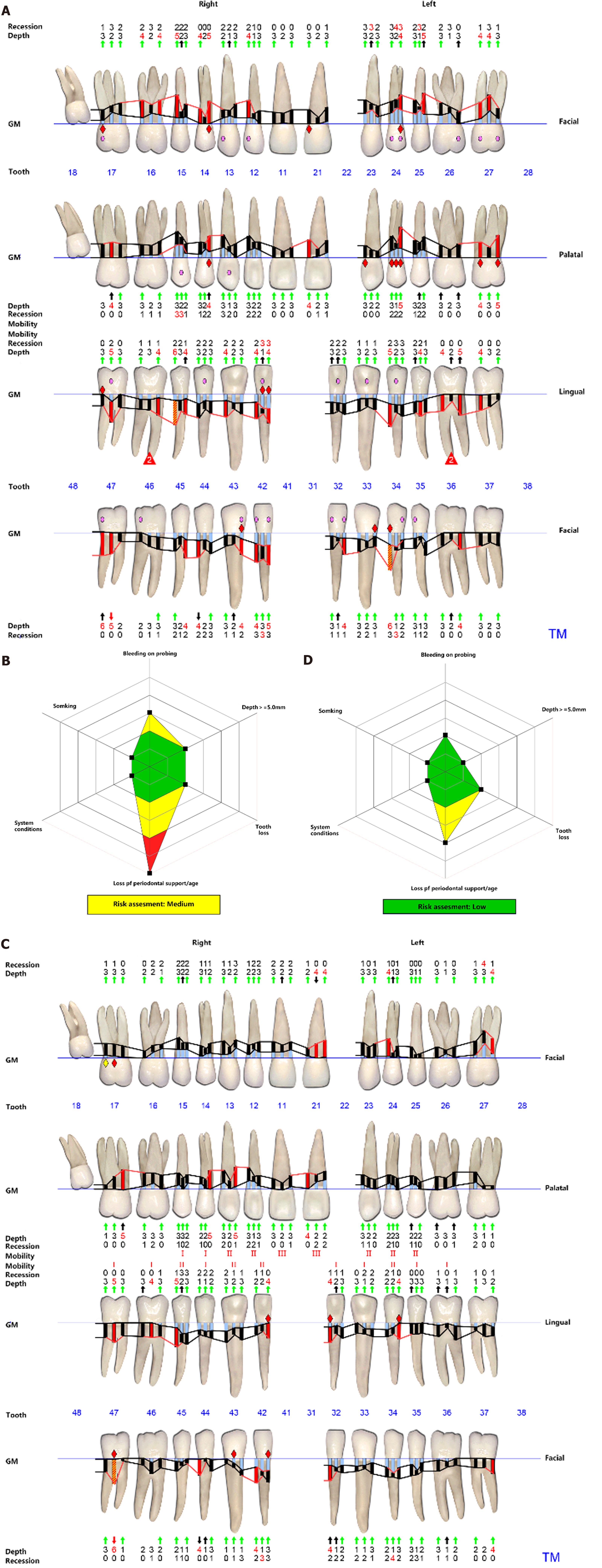
Figure 3 Periodontal charts and risk assessment.
A and C: Periodontal charts obtained after supragingival scaling and 5 mo of initial nonsurgical periodontal treatment, respectively; B and D: Respective risk assessment levels obtained using a Florida probe at the same time points. Periodontal parameters remarkably improved, although the risk assessment level remained high before orthodontic therapy. GM: Gingival margin.
Figure 4 Periodontal charts and risk assessment.
A and C: Periodontal charts obtained 1 mo after orthodontic treatment and at completion, respectively; B and D: Respective risk assessments levels obtained using a Florida probe at the same time points. Periodontal parameters continued to improve and the risk assessment level decreased from moderate to low after completion of orthodontic treatment. GM: Gingival margin.
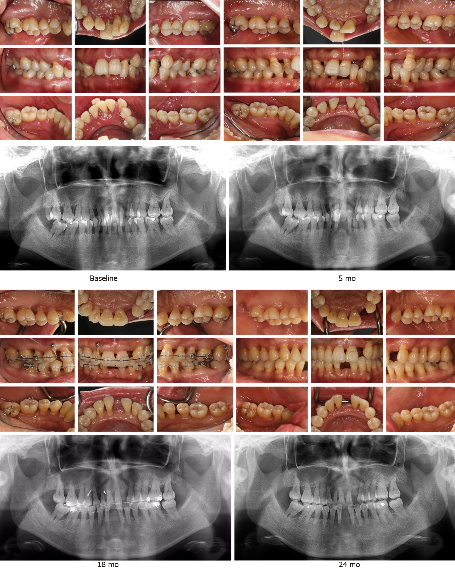
Figure 5 Intraoral and radiographic images obtained at baseline, and 5, 18, and 24 mo after treatment.
The aesthetic profile was enhanced and the radiographic bone density increased in the molar region following periodontal and orthodontic treatment. Images show the excellent therapeutic outcome following multidisciplinary treatment.
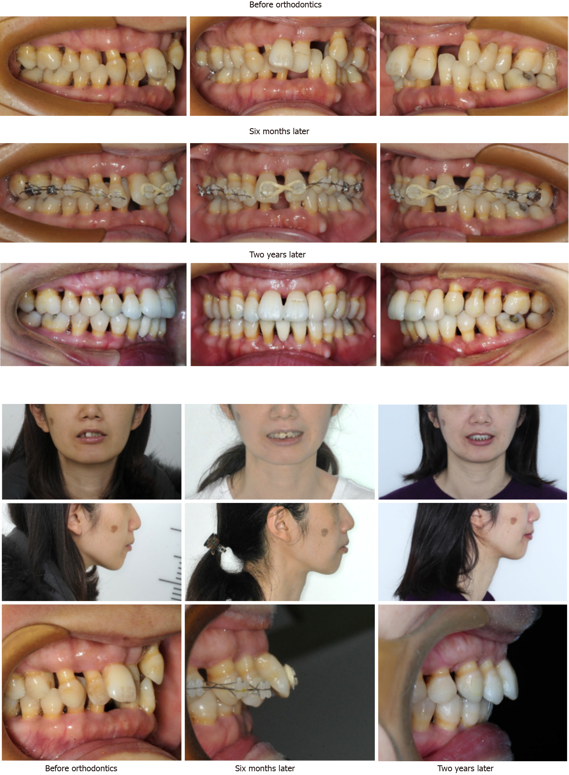
Figure 6 Intraoral and extraoral images obtained prior to, and 6 mo and 2 years following multidisciplinary treatment.
Comparative views show the significant improvement in dentition, occlusion, and the aesthetic facial profile. Note the maintenance of a healthy periodontal condition.
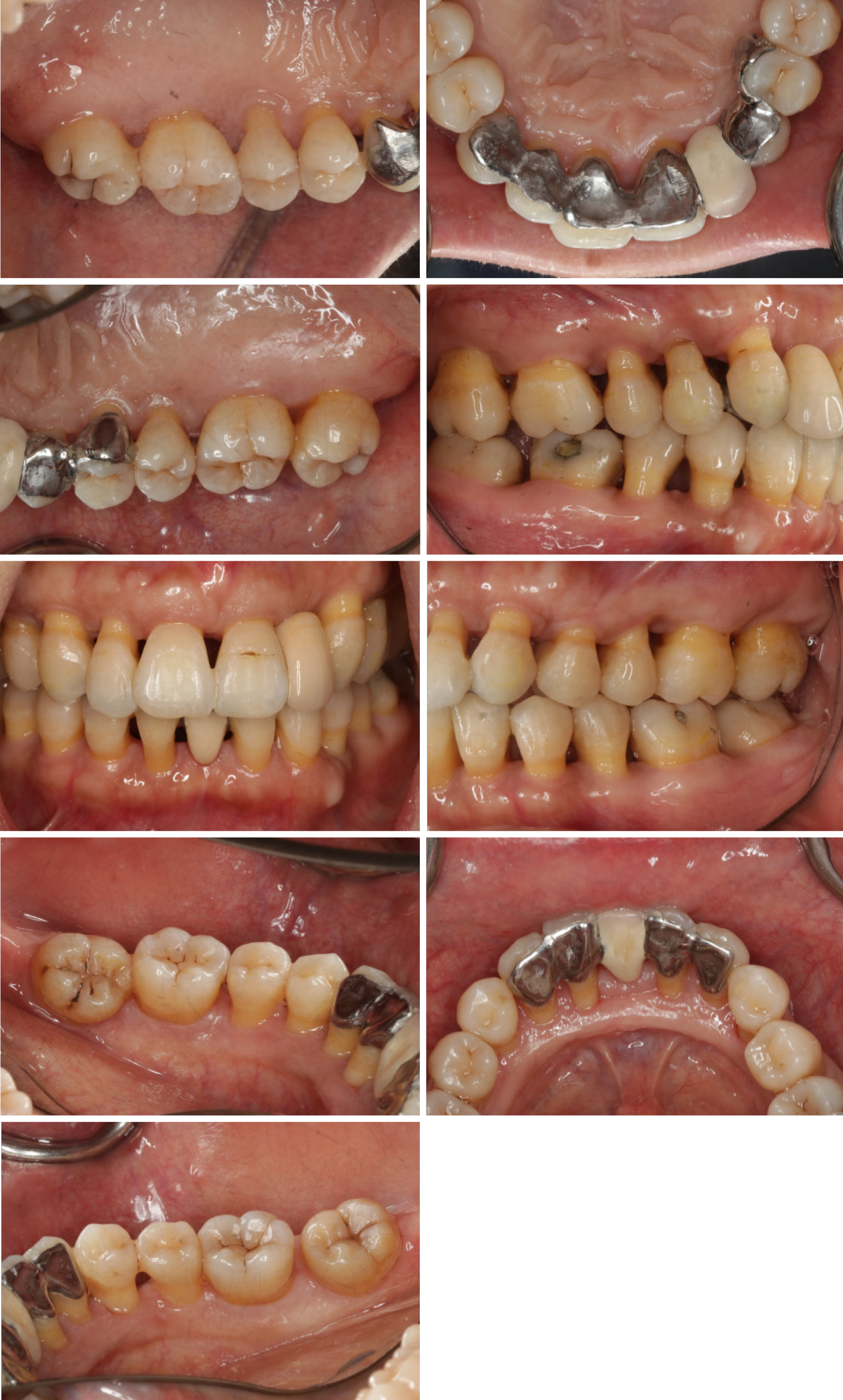
Figure 7 Intraoral images obtained after prosthodontic treatment.
A Maryland bridge was used for Nos. 13-24 and 33-43 teeth to restore teeth 22, 31, and 41. Overall, stable occlusion and a satisfactory aesthetic appearance were obtained via the comprehensive multidisciplinary approach.
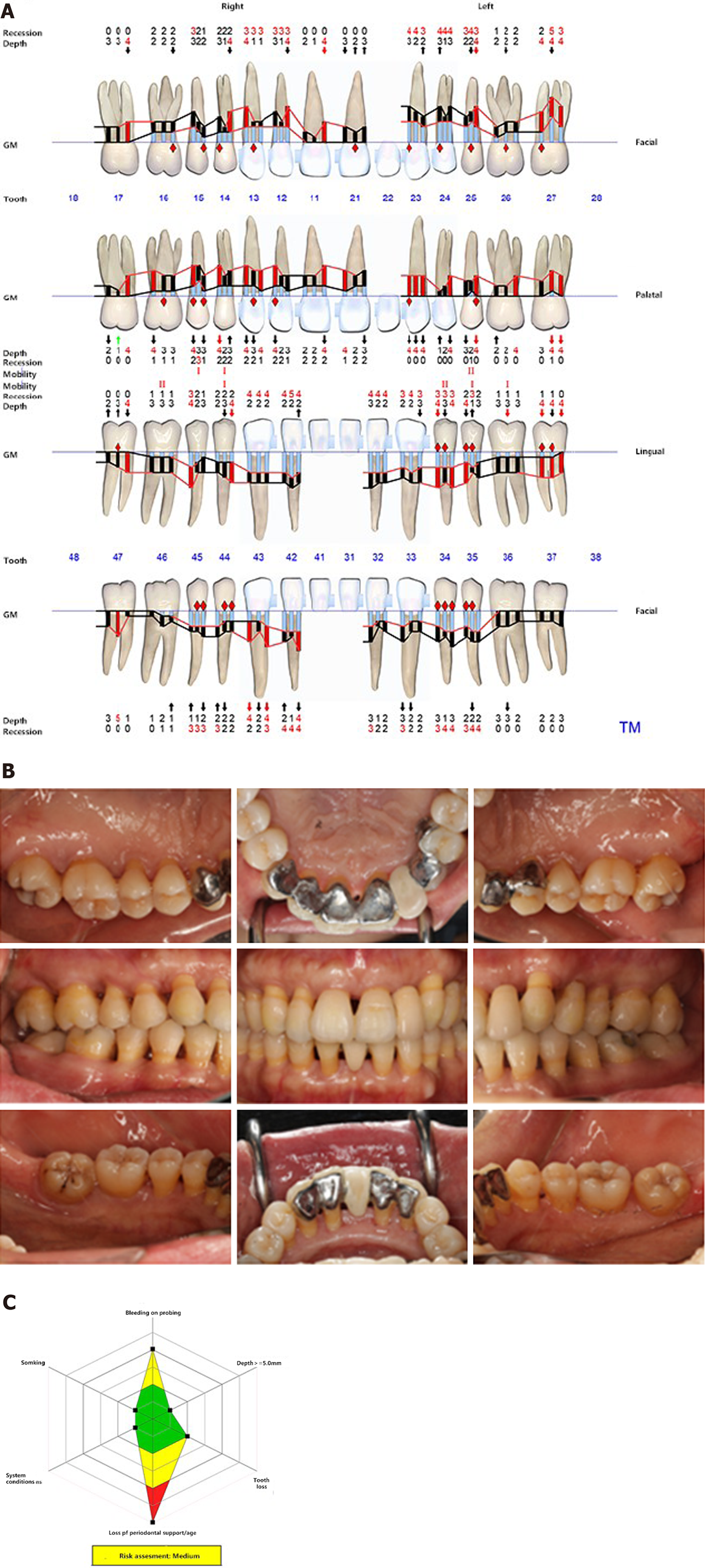
Figure 8 Periodontal condition analysis 8 mo after prosthodontic treatment (32 mo after starting treatment).
The images show the stable periodontal condition obtained as a result of the comprehensive multidisciplinary approach. A: Periodontal charts obtained 8 mo after prosthodontic treatment; B: Intraoral images taken 8 mo after prosthodontic treatment; C: Risk assessment obtained 8 mo after prosthodontic treatment.
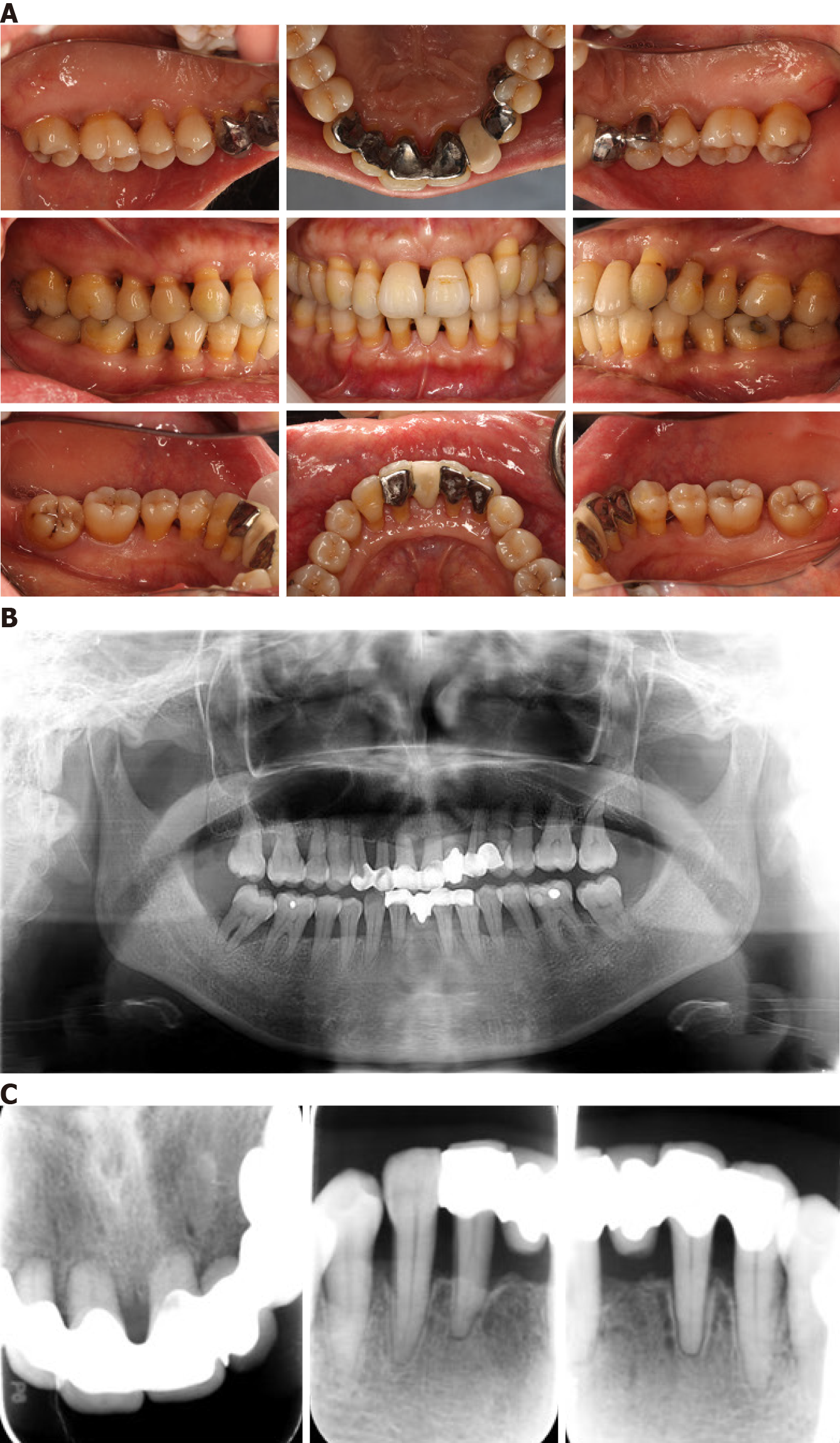
Figure 9 Periodontal condition analysis 14 mo after prosthodontic treatment (38 mo after starting treatment).
The images show the stable periodontal condition obtained as a result of the comprehensive multidisciplinary approach. A: Intraoral images taken 14 mo after prosthodontic treatment; B: Panoramic radiograph taken 14 mo after prosthodontic treatment; C: Periapical films of the upper and lower anterior teeth 14 mo after prosthodontic treatment.
- Citation: Li LJ, Yan X, Yu Q, Yan FH, Tan BC. Multidisciplinary non-surgical treatment of advanced periodontitis: A case report. World J Clin Cases 2022; 10(7): 2229-2246
- URL: https://www.wjgnet.com/2307-8960/full/v10/i7/2229.htm
- DOI: https://dx.doi.org/10.12998/wjcc.v10.i7.2229


