Copyright
©The Author(s) 2022.
World J Clin Cases. Mar 6, 2022; 10(7): 2206-2215
Published online Mar 6, 2022. doi: 10.12998/wjcc.v10.i7.2206
Published online Mar 6, 2022. doi: 10.12998/wjcc.v10.i7.2206
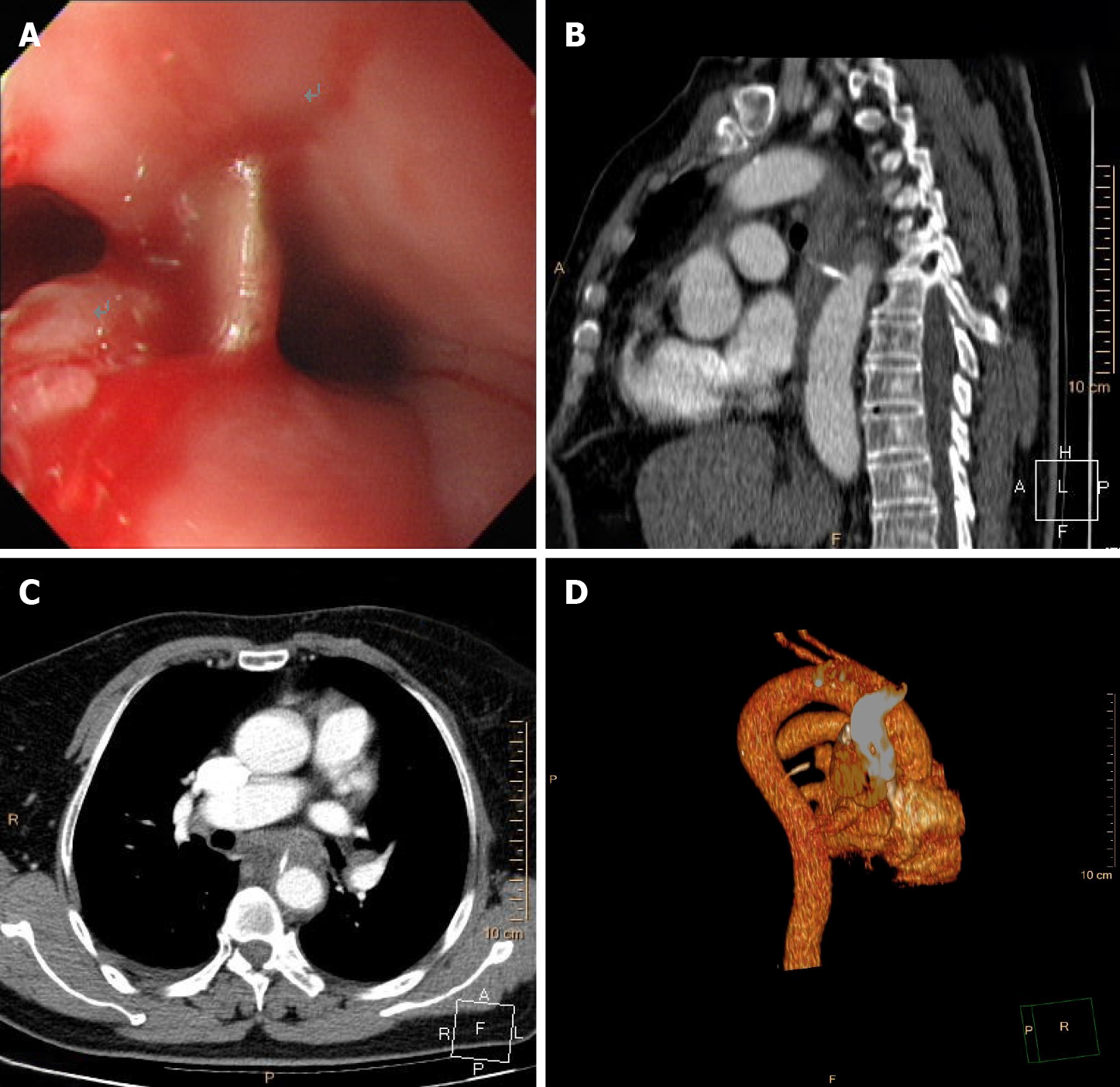
Figure 1 Esophagogastroduodenoscopy and chest computed tomography angiography.
A: Esophagogastroduodenoscopy shows a straight fishbone penetrating the wall of the esophagus, with overflowing purulent secretion, 25 cm from the incisors. The other three pictures reveal preoperative chest computed tomography angiography of the fishbone (length 2.0 cm) penetrating the wall of the esophagus and into the thoracic aorta; B: Sagittal view; C: Axial view; D: Three-dimensional reconstruction.
Figure 2 Preoperative chest computed tomography angiography shows a fishbone puncturing through the esophageal wall, tightly appressed to the descending aortic wall, accompanied by mediastinal hematoma.
A: Sagittal view; B: Axial view; C: Three-dimensional reconstruction.
Figure 3 Chest computed tomography angiography images after removal of the fishbone in case 1.
Extravasation of contrast agent presented a small nodular shadow in the thoracic aorta, and the wall of the middle esophagus was swollen and thickened. A: Sagittal view; B: Axial view; C: Three-dimensional reconstruction.
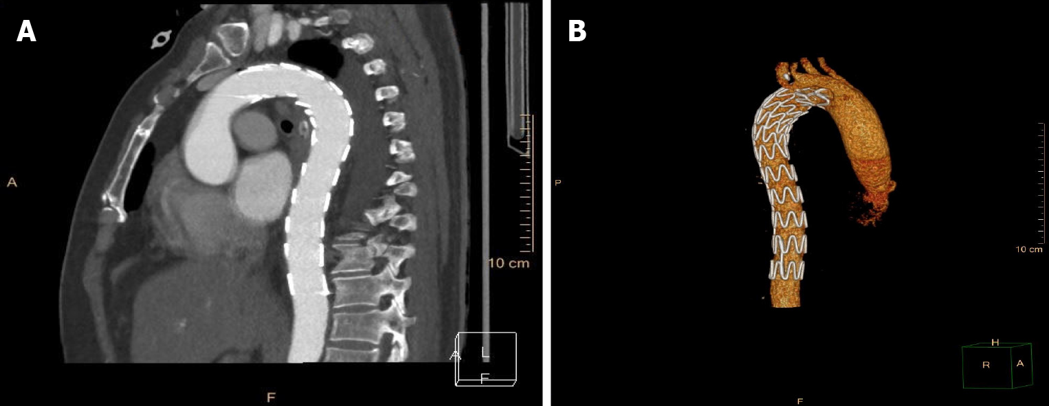
Figure 4 Chest computed tomography angiography images after endovascular stent-graft treatment in case 1.
The stent-graft was seen in the thoracic aorta without obstruction or contrast agent extravasation, and the swollen and thickened wall of the middle esophagus was significantly improved. A: Sagittal view; B: Three-dimensional reconstruction.
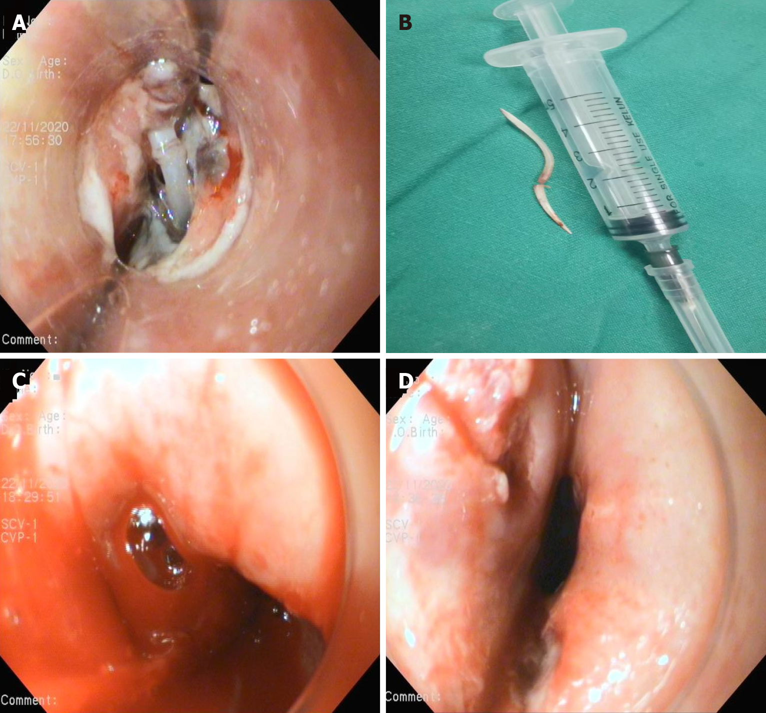
Figure 5 Esophageal view during Esophagogastroduodenoscopy.
A: Both ends of the fishbone inserted into the esophageal wall, 28 cm from the incisors; B: The endoscopically removed fishbone; C: Active blood spurting was noted in the esophageal defect after removal of the fishbone; D: Two longitudinal ulcers were present.
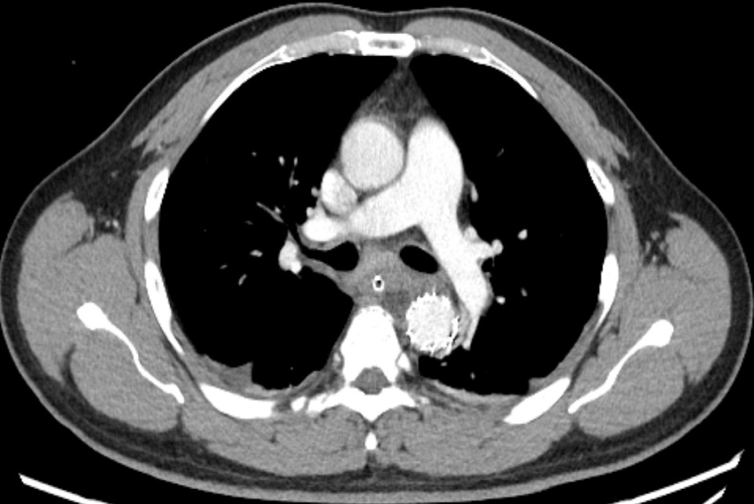
Figure 6
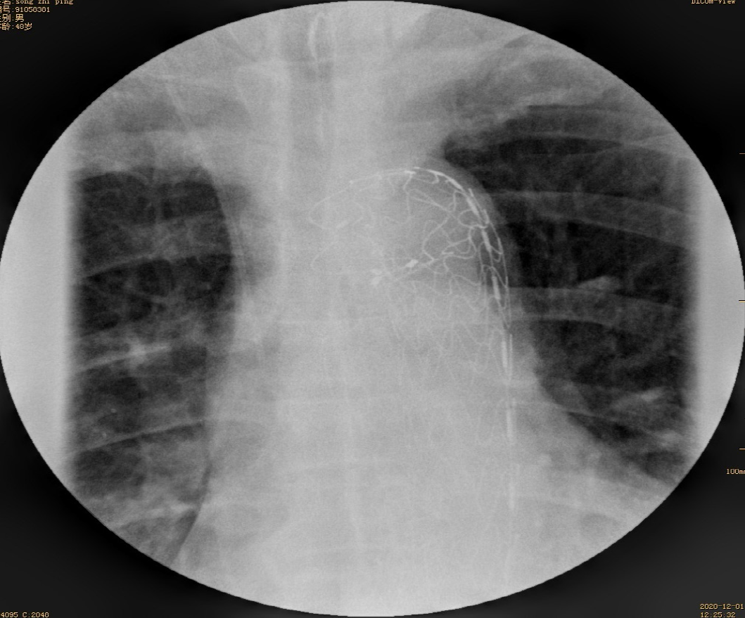
Postoperative computed tomography angiography of case 2 shows a well-positioned aortic stent graft and no contrast extravasation from the aorta.
Figure 7
Esophageal imaging with meglumine diatrizoate demonstrates no leakage.
- Citation: Gong H, Wei W, Huang Z, Hu Y, Liu XL, Hu Z. Endovascular stent-graft treatment for aortoesophageal fistula induced by an esophageal fishbone: Two cases report. World J Clin Cases 2022; 10(7): 2206-2215
- URL: https://www.wjgnet.com/2307-8960/full/v10/i7/2206.htm
- DOI: https://dx.doi.org/10.12998/wjcc.v10.i7.2206


