Copyright
©The Author(s) 2022.
World J Clin Cases. Feb 26, 2022; 10(6): 2045-2052
Published online Feb 26, 2022. doi: 10.12998/wjcc.v10.i6.2045
Published online Feb 26, 2022. doi: 10.12998/wjcc.v10.i6.2045
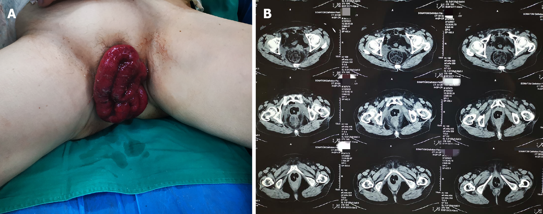
Figure 1 The small intestine had prolapsed.
A: Vaginal enterocele appears dark red in color; B: Pelvic CT suggested partial small bowel prolapse into pelvic cavity.
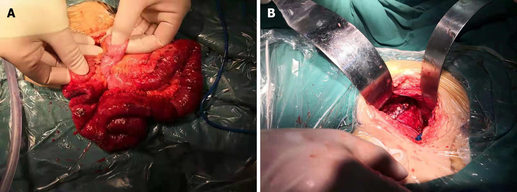
Figure 2 Open surgical exploration to ensure that the small bowel was free of mesangial torsion and necrosis.
A: During the operation, the prolapsed small intestine was still active after hot compress with warm saline solution; B: The repaired vaginal stump was reinforced to the posterior vaginal wall using a figure of eight suture.
- Citation: Liu SH, Zhang YH, Niu HT, Tian DX, Qin F, Jiao W. Vaginal enterocele after cystectomy: A case report. World J Clin Cases 2022; 10(6): 2045-2052
- URL: https://www.wjgnet.com/2307-8960/full/v10/i6/2045.htm
- DOI: https://dx.doi.org/10.12998/wjcc.v10.i6.2045


