Copyright
©The Author(s) 2022.
World J Clin Cases. Feb 26, 2022; 10(6): 2036-2044
Published online Feb 26, 2022. doi: 10.12998/wjcc.v10.i6.2036
Published online Feb 26, 2022. doi: 10.12998/wjcc.v10.i6.2036
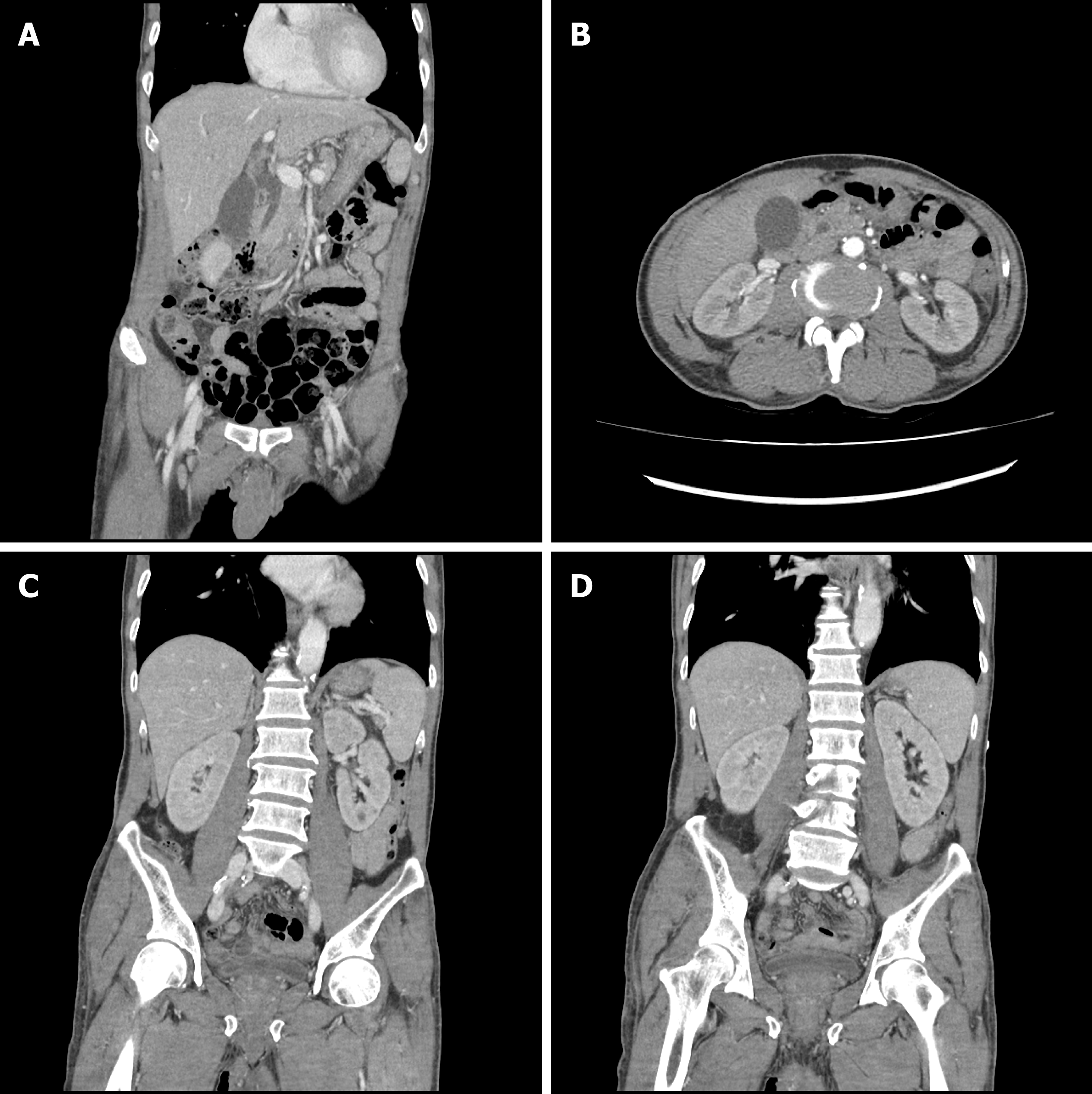
Figure 1 Computed tomography of abdomen and pelvis at the emergency department.
A: The common bile duct was mildly dilated, but it was considered as a senile change without any obvious obstructive lesion; B: Both renal parenchymal enhancements were decreased; C: Both kidney sizes and shapes were relatively normal.
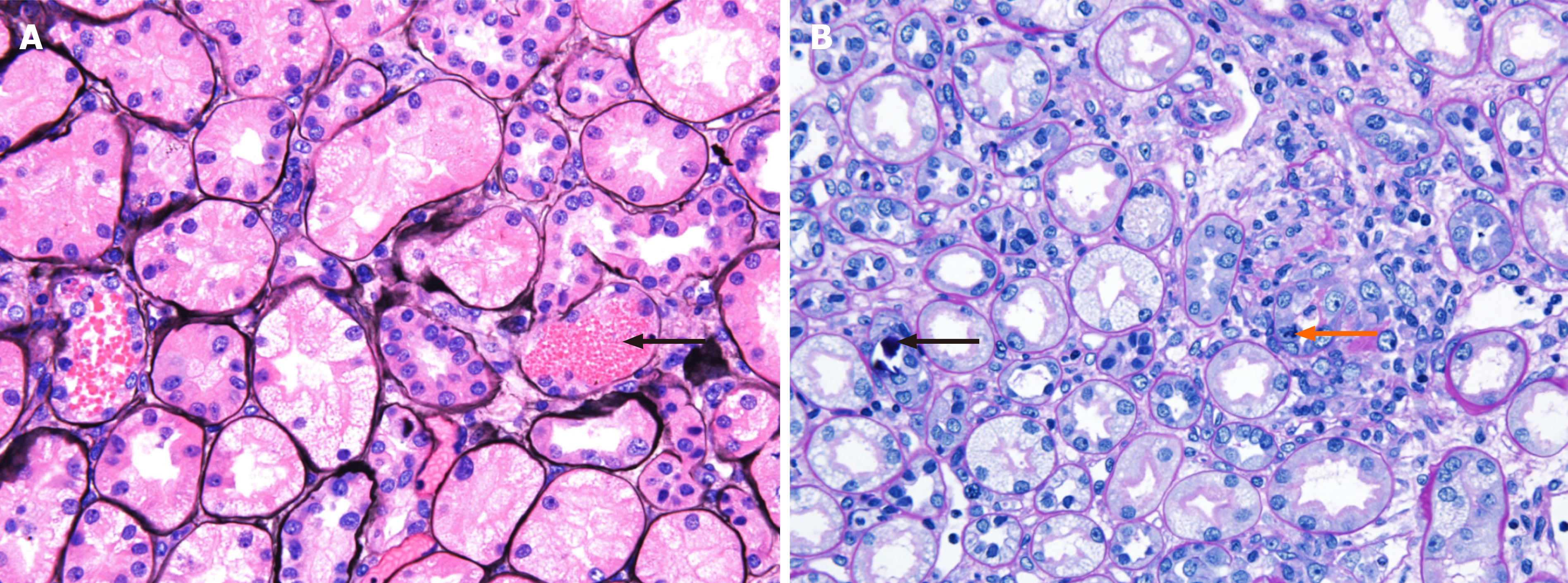
Figure 2 Light micrographs of renal biopsy.
A: The tubules show vacuolated degeneration with some red blood cells, granular materials (black arrow) (methenamine silver stain, × 400); B: The tubules show calcium concretions (black arrow) in tubular lumina and mitosis (orange arrow) (periodic acid-Schiff stain, × 400).
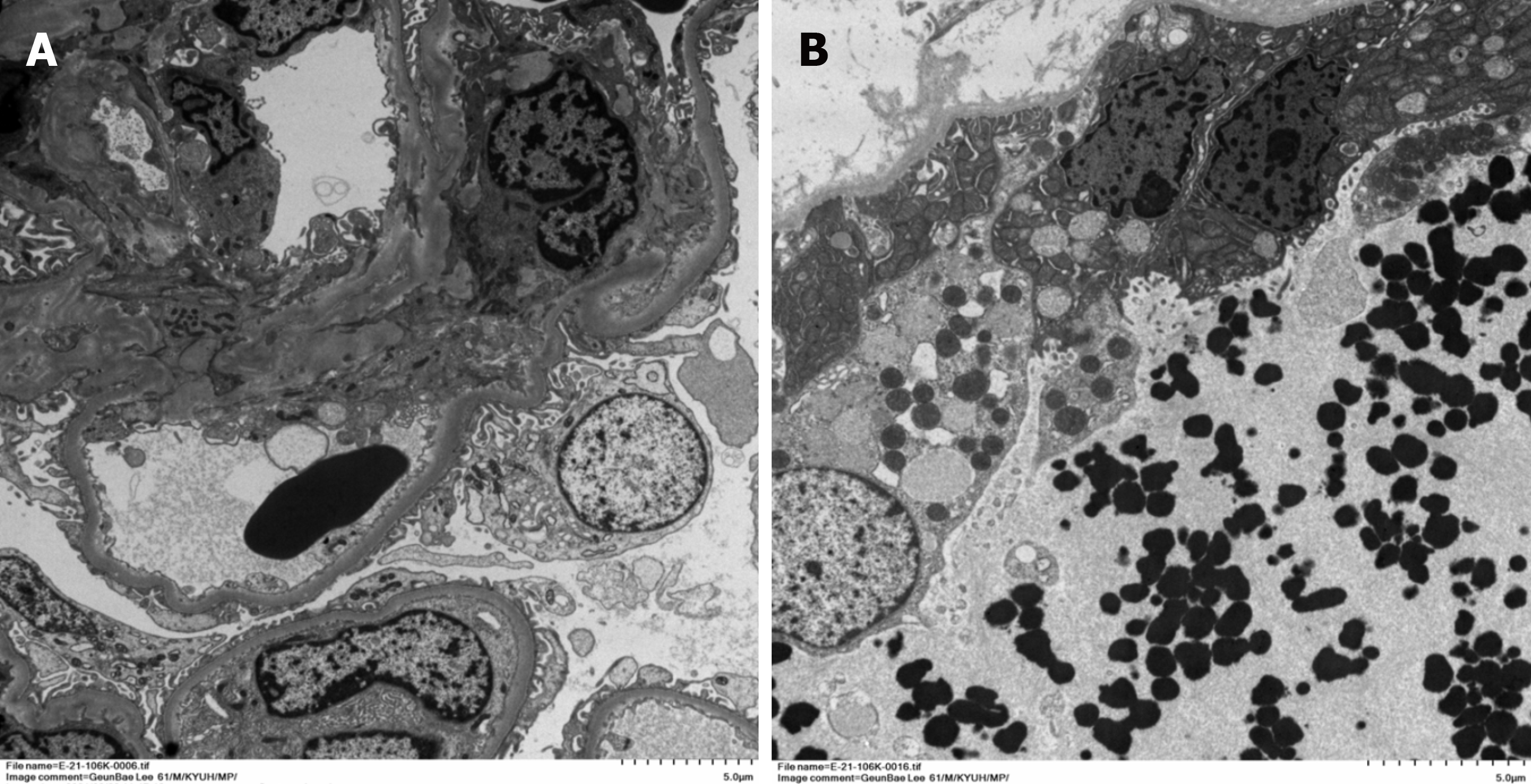
Figure 3 Electron micrographs of renal biopsy.
A: The glomerulus is well preserved with focal foot process effacement at 10% of the external capillary surface (original magnification, × 1000); B: Some distal tubules show vacuolar degenerative change with electron dense granular in distal tubular lumen (original magnification, × 1200).
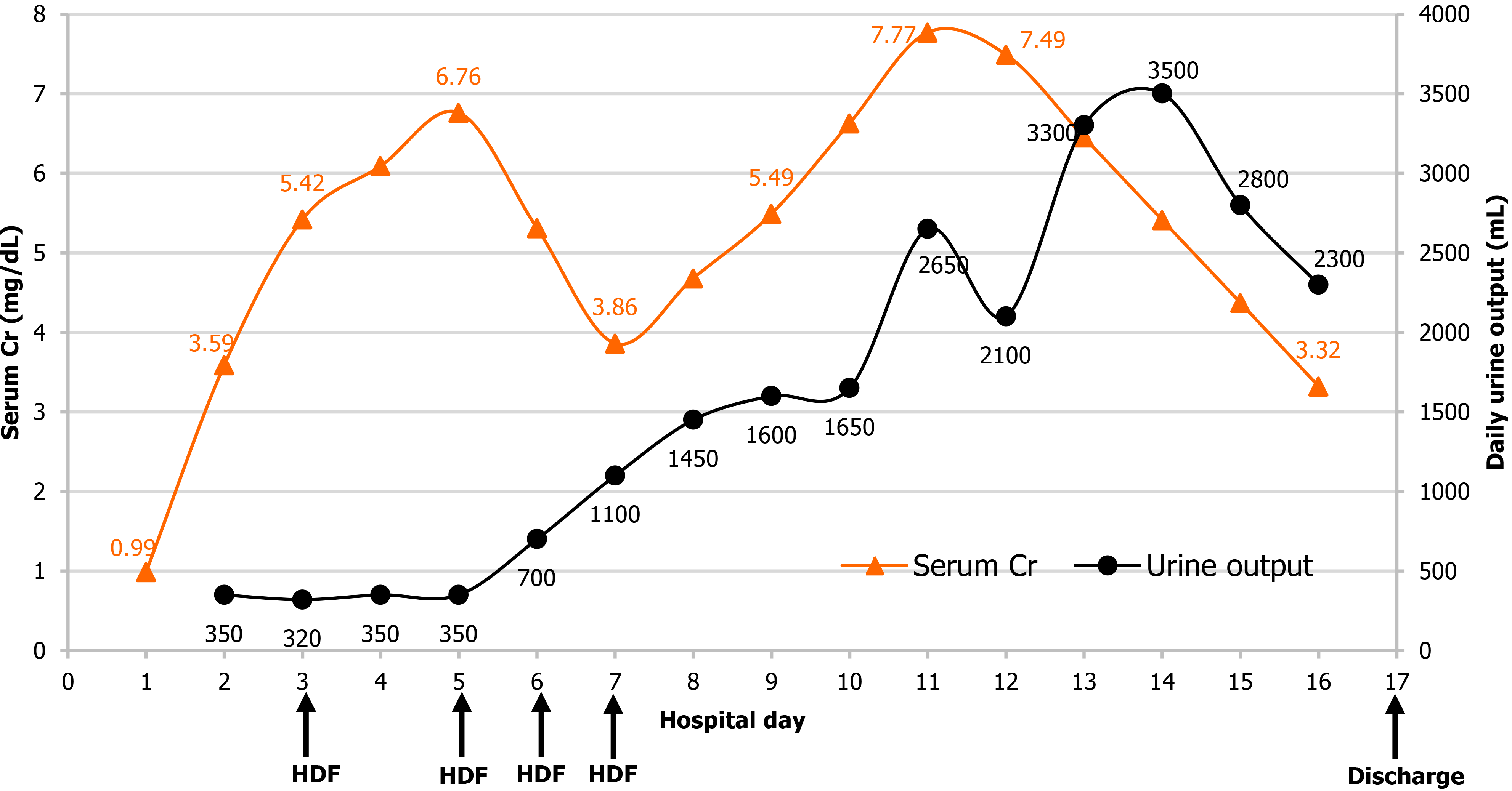
Figure 4 Changes in the serum creatinine level and urine output during hospitalization.
On the 2nd day of hospitalization, the serum creatinine (Cr) level increased while the urine output decreased. A total of four hemodiafiltration sessions were performed, and urine output gradually increased from the 7th day of hospitalization. The serum Cr level began to decrease from the 12th day of hospitalization, and the patient was discharged on the 17th day of hospitalization without any sequelae. HDF: Hemodiafiltration; Cr: Creatinine.
- Citation: Park S, Ryu HS, Lee JK, Park SS, Kwon SJ, Hwang WM, Yun SR, Park MH, Park Y. Acute kidney injury due to intravenous detergent poisoning: A case report. World J Clin Cases 2022; 10(6): 2036-2044
- URL: https://www.wjgnet.com/2307-8960/full/v10/i6/2036.htm
- DOI: https://dx.doi.org/10.12998/wjcc.v10.i6.2036


