Copyright
©The Author(s) 2022.
World J Clin Cases. Feb 26, 2022; 10(6): 2030-2035
Published online Feb 26, 2022. doi: 10.12998/wjcc.v10.i6.2030
Published online Feb 26, 2022. doi: 10.12998/wjcc.v10.i6.2030
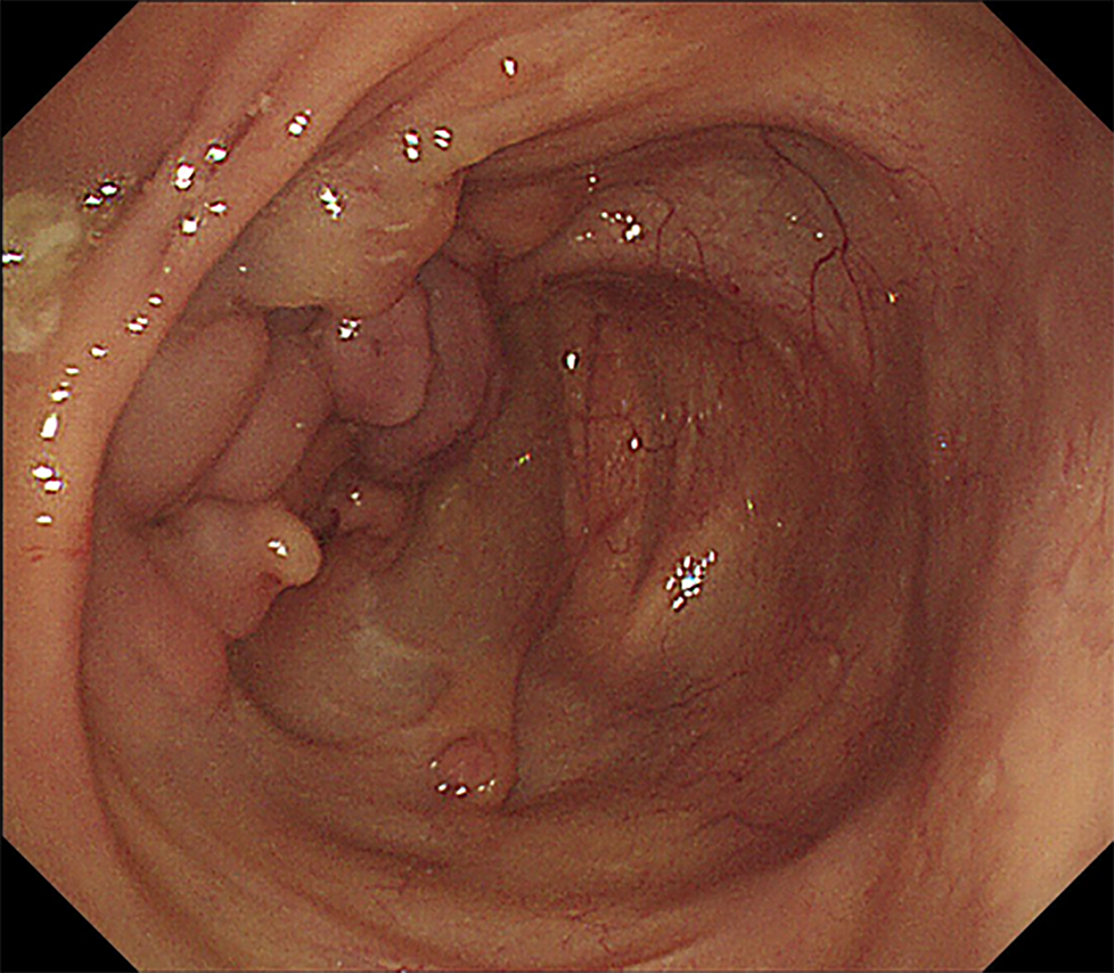
Figure 1 Colonoscopy showing deformation, mucosal hyperplasia and multiple deep ulcers in the ileocecal region.
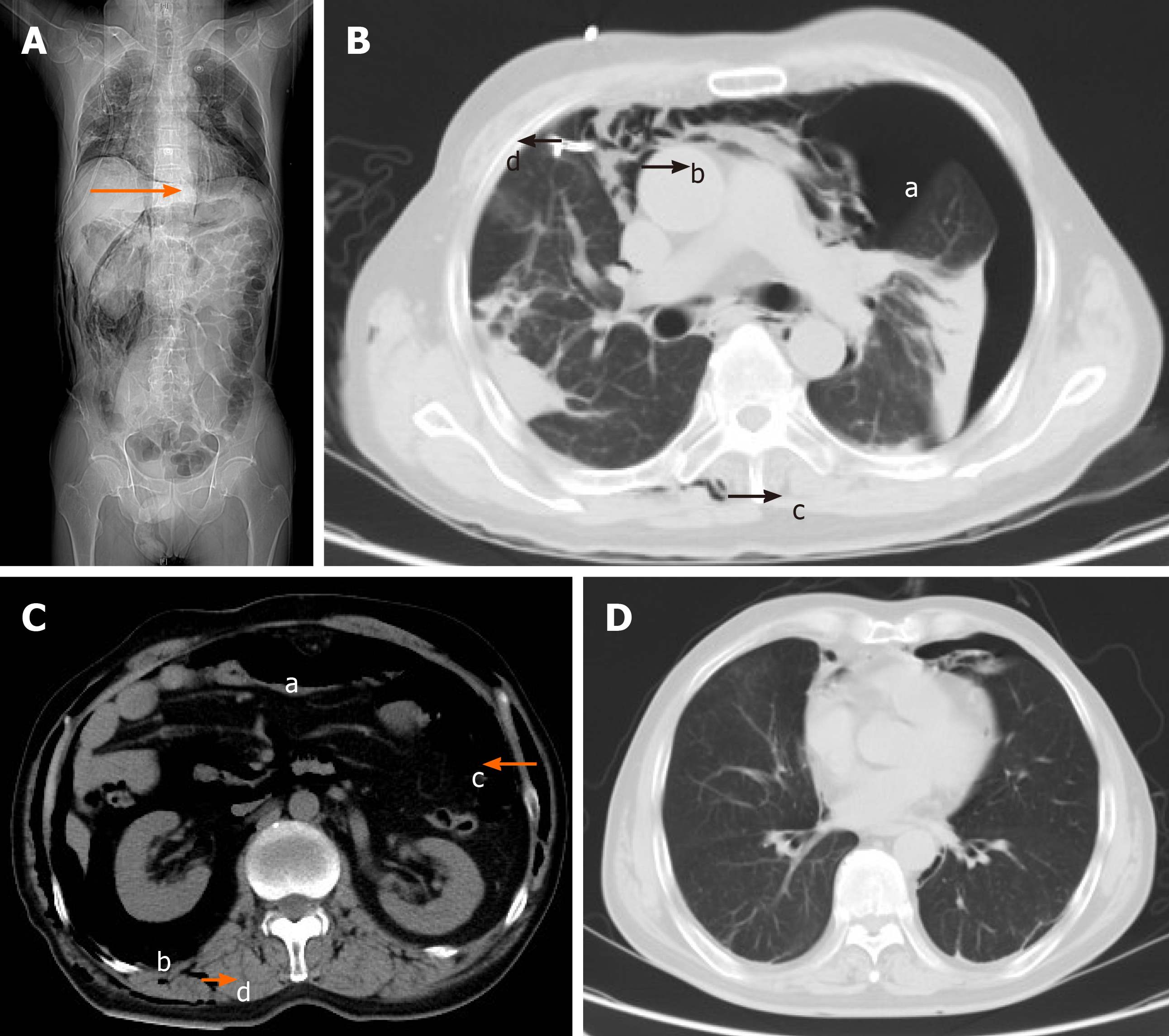
Figure 2 Computed tomography.
A: Computed tomography (CT) scan showing massive free air and right chest drain tube (a1, arrow); B: CT scan showing bilateral pneumothorax (b1), pneumomediastinum (b2), subcutaneous emphysema of the back (b3) and right chest drain tube (b4); C: CT scan showing pneumoperitoneum (c1), pneumoretroperitoneum (c2) and subcutaneous emphysema of the abdomen (c3) and back (c4); D: CT scan showing significant improvement of bilateral pneumothorax and pneumomediastinum.
- Citation: Mu T, Feng H. Bilateral pneumothorax and pneumomediastinum during colonoscopy in a patient with intestinal Behcet’s disease: A case report. World J Clin Cases 2022; 10(6): 2030-2035
- URL: https://www.wjgnet.com/2307-8960/full/v10/i6/2030.htm
- DOI: https://dx.doi.org/10.12998/wjcc.v10.i6.2030


