Copyright
©The Author(s) 2022.
World J Clin Cases. Feb 26, 2022; 10(6): 2015-2022
Published online Feb 26, 2022. doi: 10.12998/wjcc.v10.i6.2015
Published online Feb 26, 2022. doi: 10.12998/wjcc.v10.i6.2015
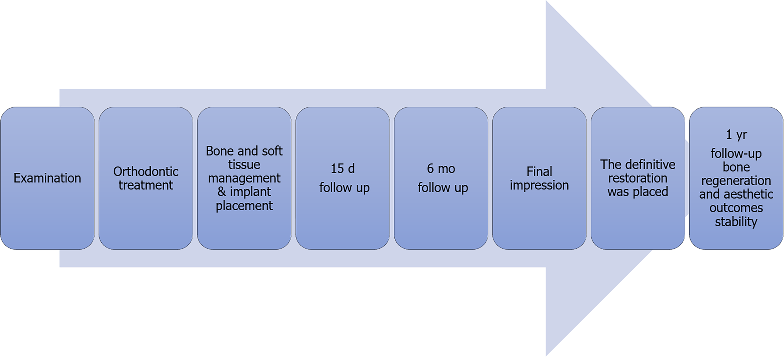
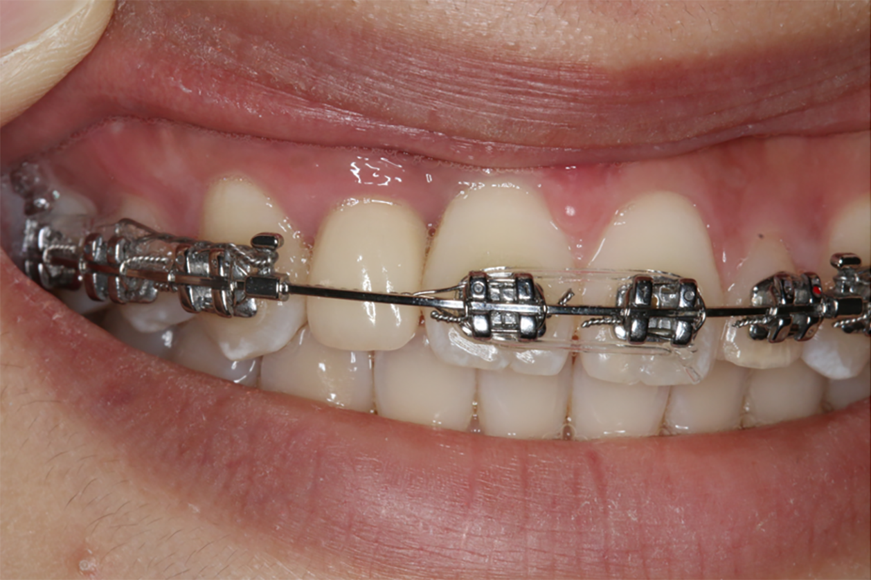
Figure 1 Flow chart timeline of the treatment plan.
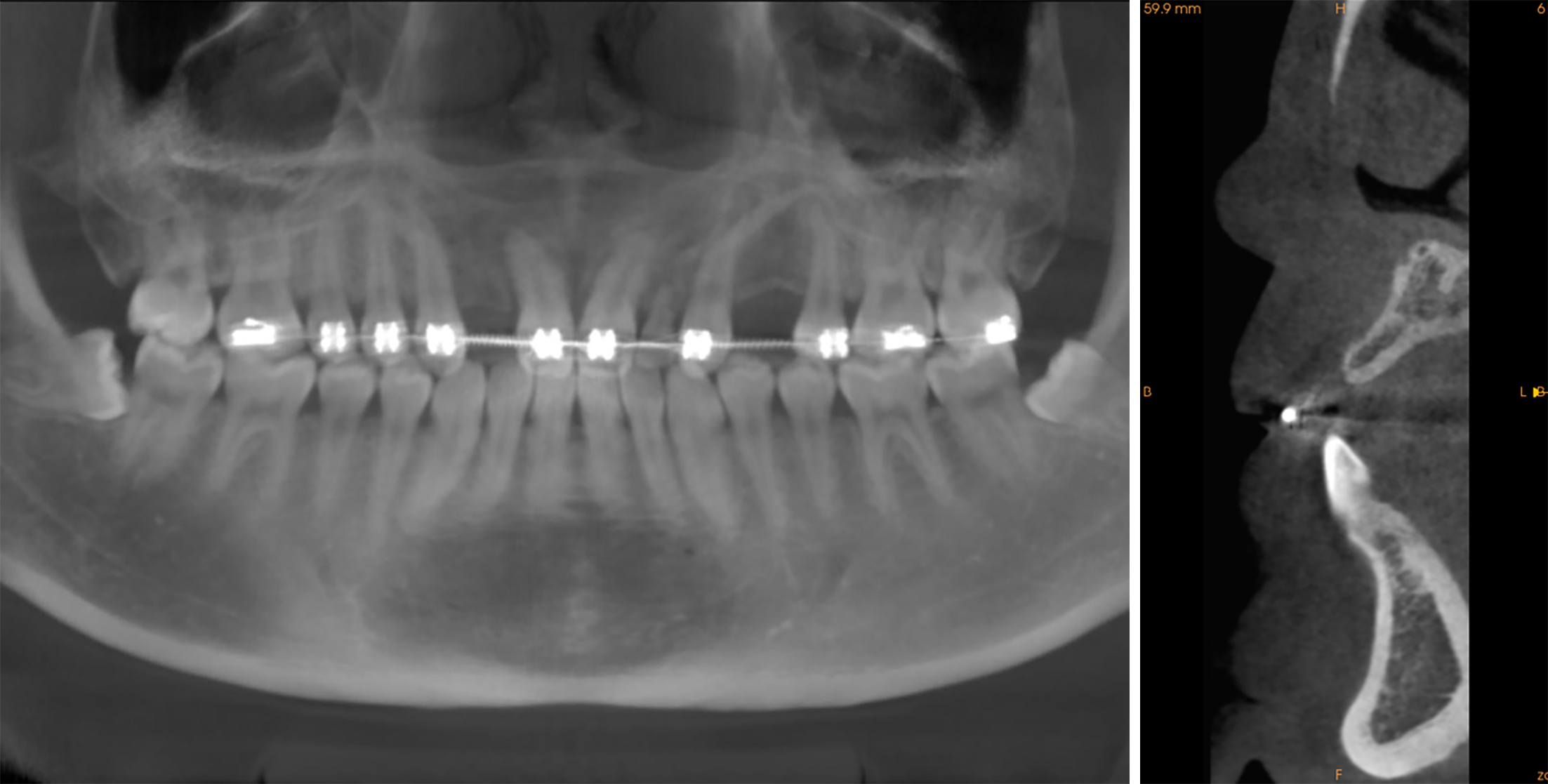
Figure 2 Cone-beam computed tomography revealed considerable alveolar bone loss.
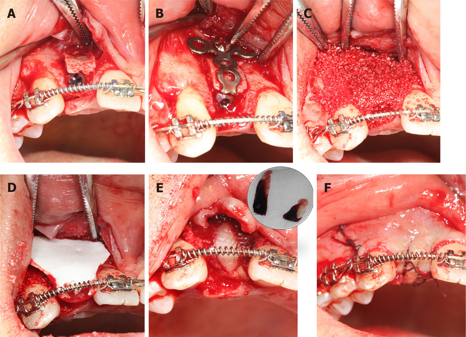
Figure 3 The surgical procedure.
A: The implant was placed at the recipient cite #12 after the labial bone plate displacement; B: A T-type titanium plate was used to fix the bone block; C: The mixture of bone grafts and platelet-rich fibrin (PRF) clot covered the T-type titanium plate and the socket walls; D: Resorbable membrane covered the bone grafts; E: PRF membrane covered the resorbable membrane and alveolar crest; F: The wound was non-tightly sutured.
Figure 4 Intraoral condition at the 15 d follow-up visit: The vascularization of soft tissue was visible.
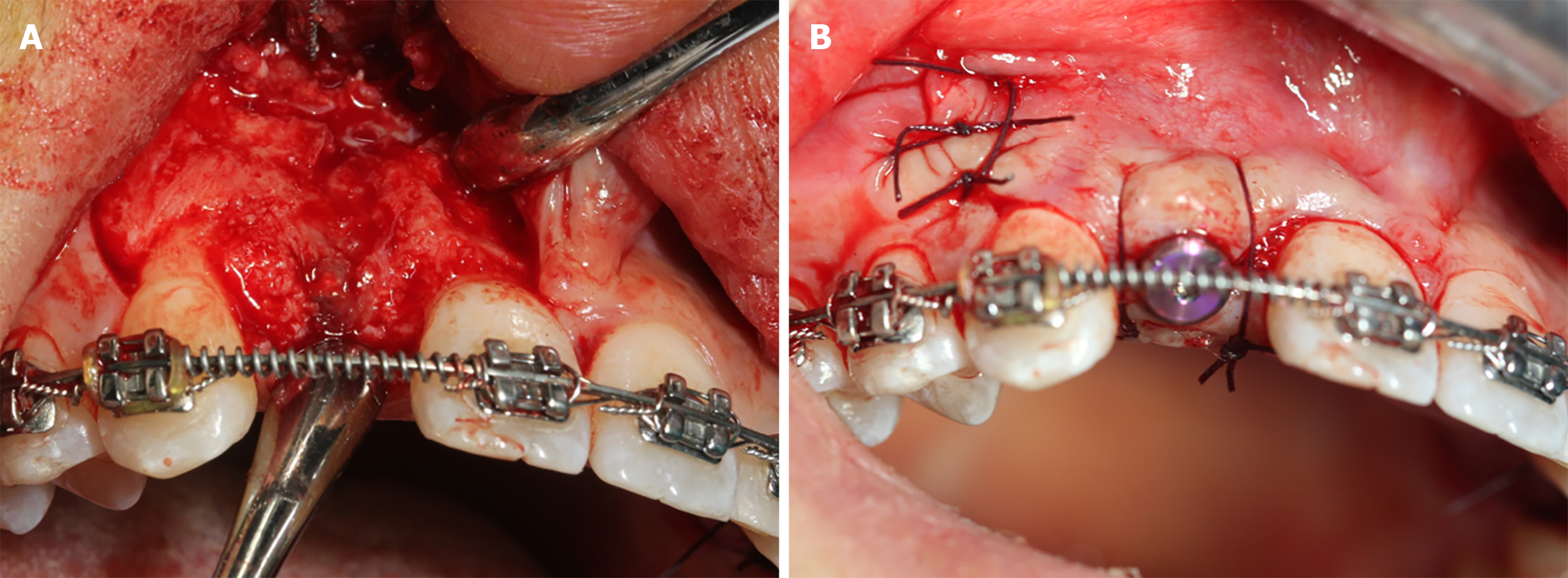
Figure 5 Second stage surgery.
A: The implant was surrounded by bone and the titanium plate was covered by the new bone; B: The incision was non-tightly sutured.
Figure 6 The definitive restoration.
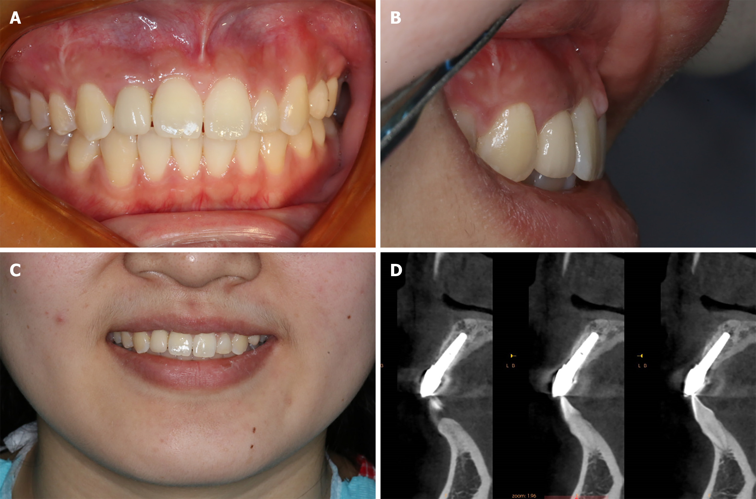
Figure 7 Assessments at 1 year after surgery.
A-C: Intraoral condition; D: Cone-beam computed tomography image showing the stable bone around implant.
- Citation: Zhang TS, Mudalal M, Ren SC, Zhou YM. Implant site development using titanium plate and platelet-rich fibrin for congenitally missed maxillary lateral incisors: A case report. World J Clin Cases 2022; 10(6): 2015-2022
- URL: https://www.wjgnet.com/2307-8960/full/v10/i6/2015.htm
- DOI: https://dx.doi.org/10.12998/wjcc.v10.i6.2015


