Copyright
©The Author(s) 2022.
World J Clin Cases. Feb 26, 2022; 10(6): 1991-1997
Published online Feb 26, 2022. doi: 10.12998/wjcc.v10.i6.1991
Published online Feb 26, 2022. doi: 10.12998/wjcc.v10.i6.1991
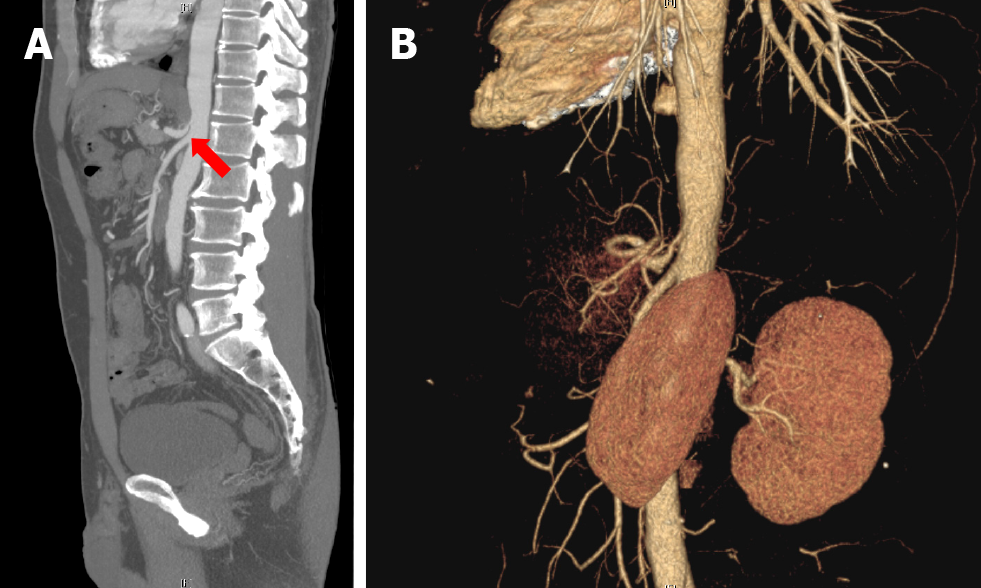
Figure 1 Imaging examinations of case 1.
A: Computed tomography (CT) shows the celiac trunk originating from the upper abdominal aorta rather than in the normal case; B: CT angiography shows narrowing of the orfice of the celiac trunk due to compression by the diaphragmatic crus.
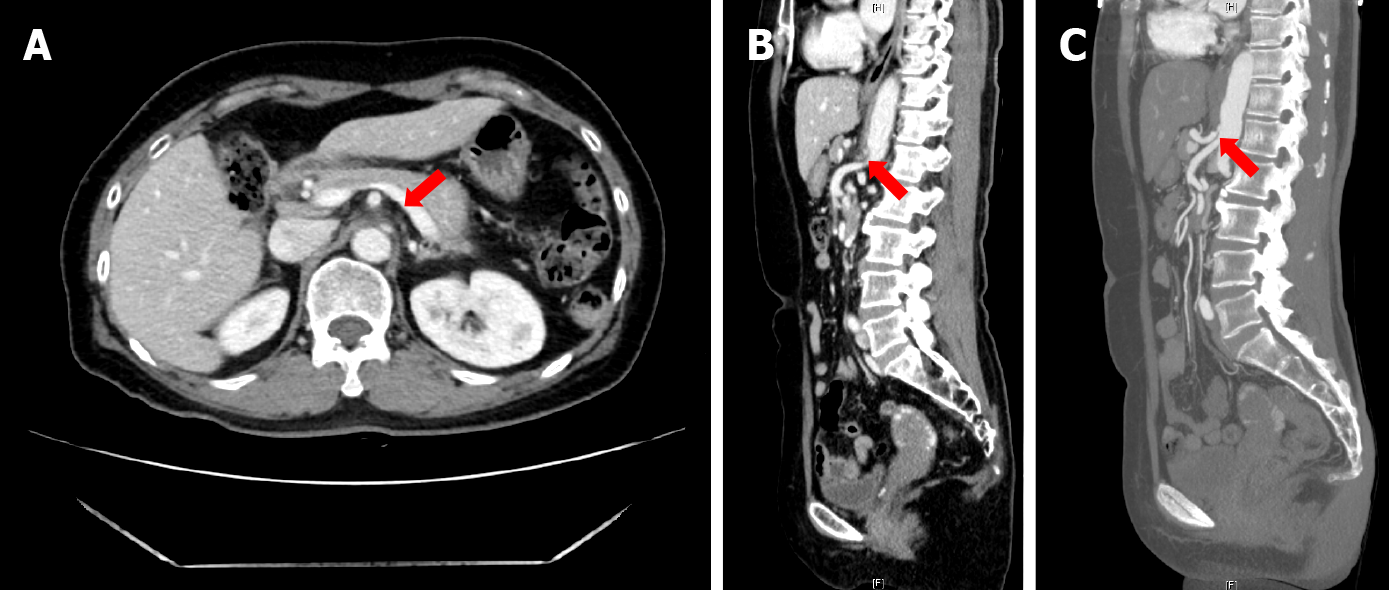
Figure 2 Computed tomography shows the proximal celiac axis narrowing with collateral vessel formation.
A: Axial view; B: Sagittal view; C: Computed tomography angiography confirmed the diagnosis.
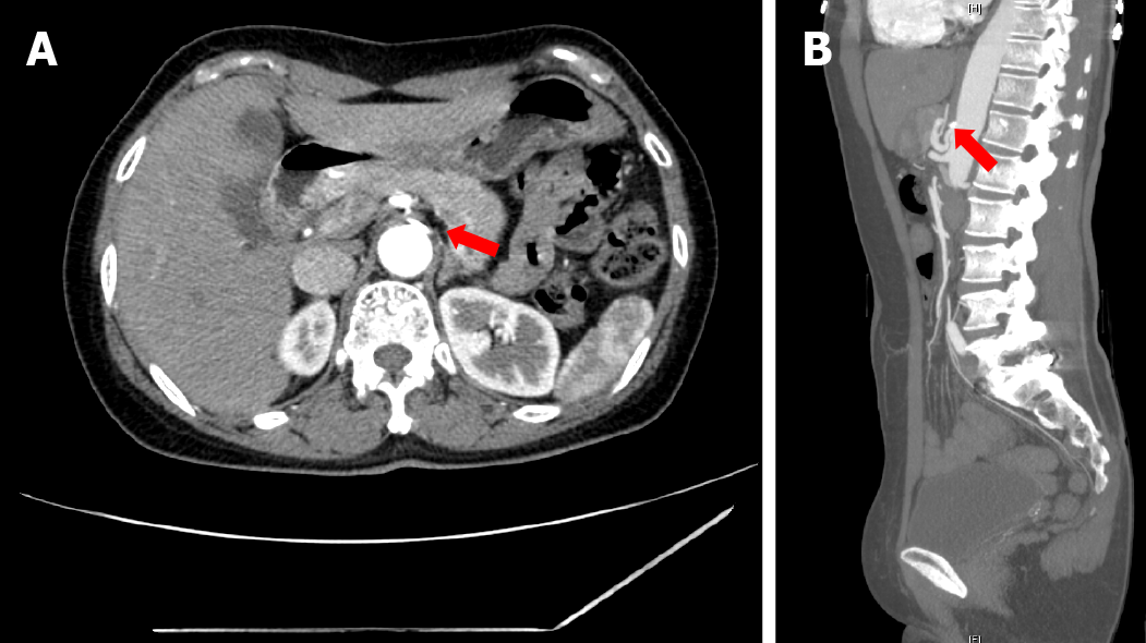
Figure 3 Computed tomography angiography shows mild luminal narrowing at the orifice of the celiac trunk with adjacent abdominal aortic calcification.
A: Axial view; B: Sagittal view.
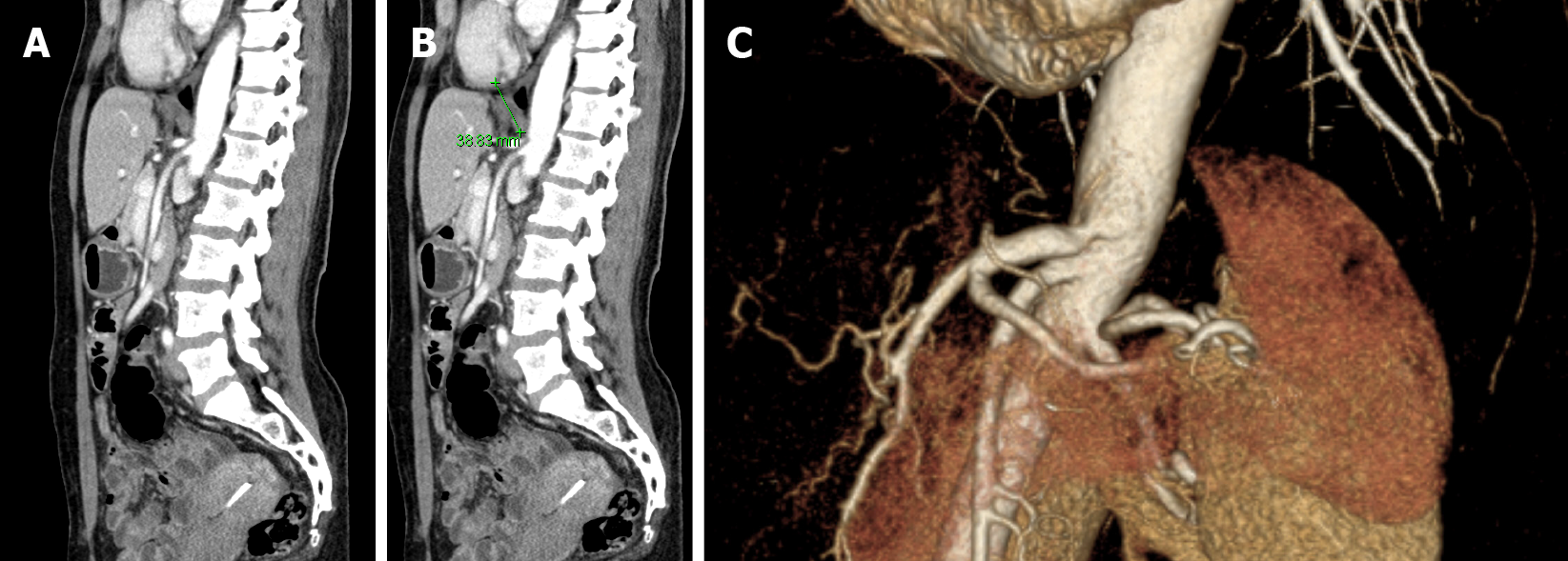
Figure 4 Imaging examinations of case 4.
A: Computed tomography (CT) showing the surrounding celiac trunk, where post-stenotic dilatation is seen; B: The median arcuate ligamentum compresses the narrowed area; C: CT angiography showing that the celiac trunk orifice is significantly narrowed, and post-stenotic dilatation is also seen.
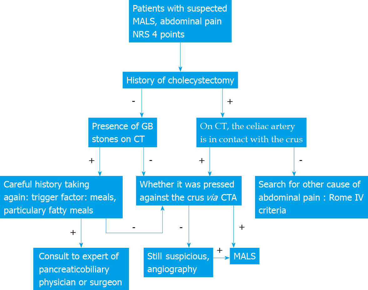
Figure 5 Algorithm for median arcuate ligamentum syndrome diagnosis through imaging modalities and clinical history.
MALS: Median arcuate ligamentum syndrome; NRS: Numeric pain rating scale; GB: Gallbladder; CT: Computed tomography; CTA: Computed tomography angiography.
- Citation: Kim JE, Rhee PL. Median arcuate ligamentum syndrome: Four case reports. World J Clin Cases 2022; 10(6): 1991-1997
- URL: https://www.wjgnet.com/2307-8960/full/v10/i6/1991.htm
- DOI: https://dx.doi.org/10.12998/wjcc.v10.i6.1991


