Copyright
©The Author(s) 2022.
World J Clin Cases. Feb 26, 2022; 10(6): 1973-1980
Published online Feb 26, 2022. doi: 10.12998/wjcc.v10.i6.1973
Published online Feb 26, 2022. doi: 10.12998/wjcc.v10.i6.1973
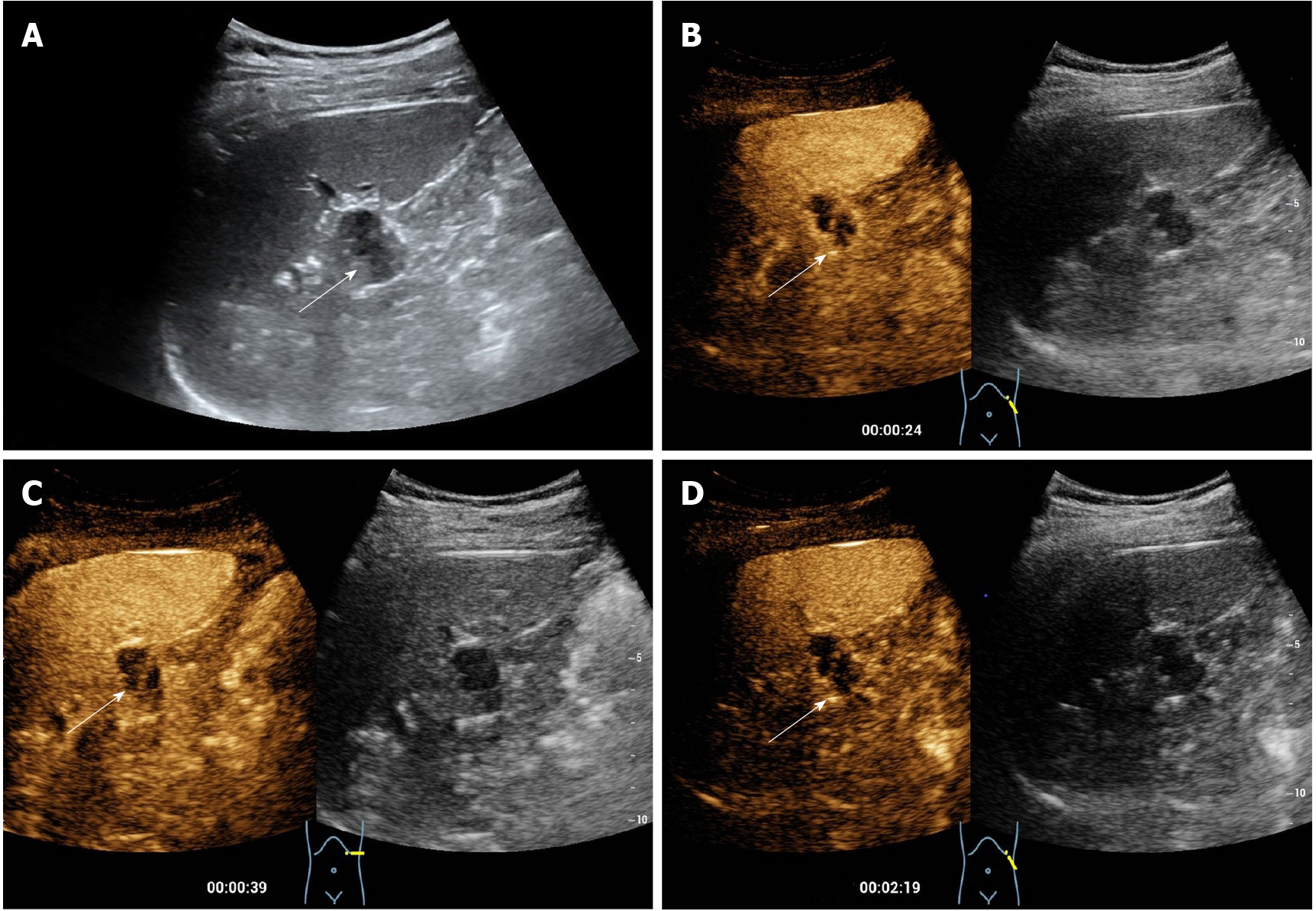
Figure 1 Pre-contrast and contrast enhanced ultrasound of the pancreatic lesion.
A: A complicated cystic lesion (arrow) measuring 2 cm was detected in the tail of the pancreas by grayscale ultrasound in a 32-year-old male patient; B: Peripheral nodular and internal septal isoenhancement (arrow) in the arterial phase was shown on contrast-enhanced ultrasound; C and D: The enhanced part of the lesion exhibited mild hyperenhancement in the early venous phase without definite washout in the late venous phase. The cystic component did not show any enhancement through either phase.
Figure 2 Pre-operative computed tomography scan of the pancreatic lesion.
A: A slightly low-density nodule measuring 2.2 cm (arrow) was found in the tail of the pancreas on unenhanced computed tomography (CT); B and C: Septa were faintly visible whereas no salient enhancement was presented within the lesion (arrows) in either the arterial or the venous phases on axial contrast-enhanced CT.
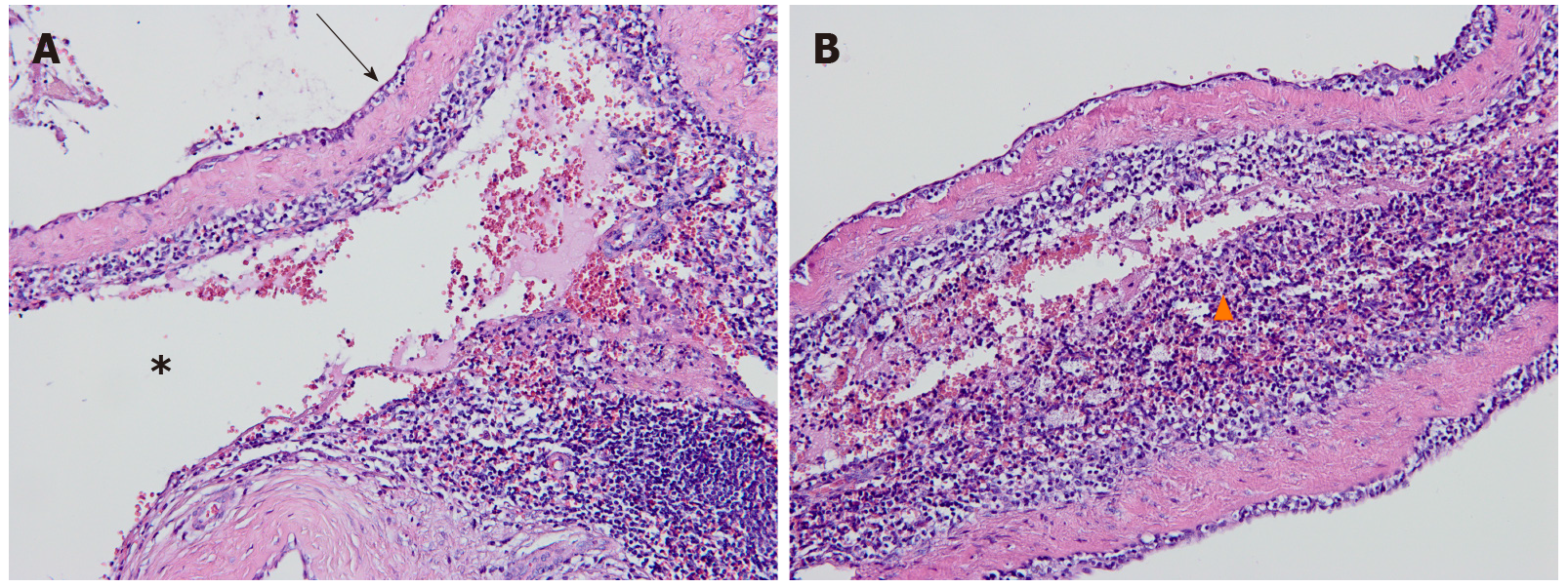
Figure 3 Hematoxylin-eosin staining of the cavernous hemangioma arising from the intrapancreatic accessory spleen.
A: Large dilated vascular spaces (asterisk) separated by fibrous septa and endothelial cells (arrows) lining on the surface of the vascular spaces were observed in the intermediate-power view (original magnification, 200×); B: A high-powered photomicrograph (original magnification, 400×) illustrated splenic tissues (triangles) adjacent to the vascular spaces.
- Citation: Huang JY, Yang R, Li JW, Lu Q, Luo Y. Cavernous hemangioma of an intrapancreatic accessory spleen mimicking a pancreatic tumor: A case report. World J Clin Cases 2022; 10(6): 1973-1980
- URL: https://www.wjgnet.com/2307-8960/full/v10/i6/1973.htm
- DOI: https://dx.doi.org/10.12998/wjcc.v10.i6.1973


