Copyright
©The Author(s) 2022.
World J Clin Cases. Feb 26, 2022; 10(6): 1966-1972
Published online Feb 26, 2022. doi: 10.12998/wjcc.v10.i6.1966
Published online Feb 26, 2022. doi: 10.12998/wjcc.v10.i6.1966
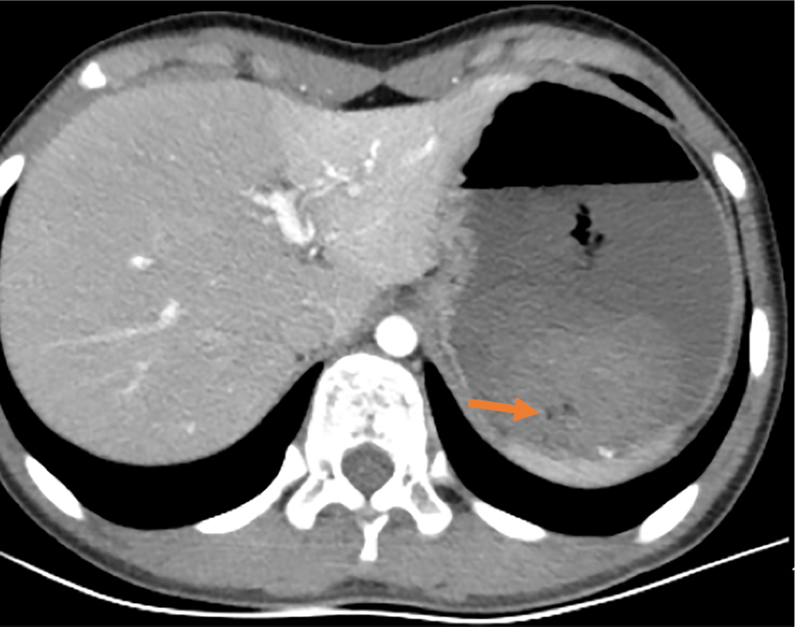
Figure 1 Abdominal enhanced computed tomography.
It showed that the stomach was visibly dilated and filled with fluid, with blood clots visible. The arrow indicates the blood clot.
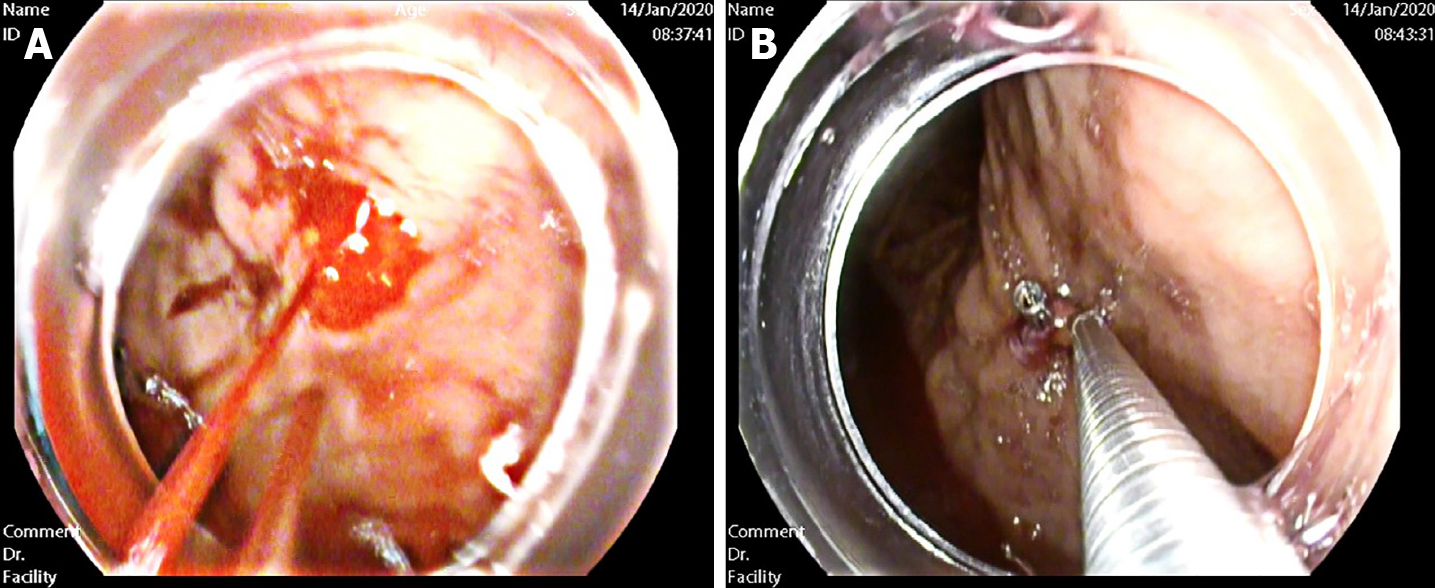
Figure 2 Endoscopic exam.
A: Endoscopic changes before hemostasis. The presence of an actively bleeding protruding vessel in the posterior wall of the body of the stomach; B: Endoscopic hemostasis. Electrocoagulation lasted 2-3 s and the power used in electrocoagulation was 40 W. Two endoscopic hemoclips were applied, which achieved full control of the bleeding.
Figure 3 Endoscopic appearance of other parts after hemostasis.
A and B: Duodenal mucosa showed no other bleeding spots, erosions, or ulcers; C: Gastric antrum was normal.
- Citation: Chen Y, Sun M, Teng X. Therapeutic endoscopy of a Dieulafoy lesion in a 10-year-old girl: A case report. World J Clin Cases 2022; 10(6): 1966-1972
- URL: https://www.wjgnet.com/2307-8960/full/v10/i6/1966.htm
- DOI: https://dx.doi.org/10.12998/wjcc.v10.i6.1966


