Copyright
©The Author(s) 2022.
World J Clin Cases. Feb 26, 2022; 10(6): 1929-1936
Published online Feb 26, 2022. doi: 10.12998/wjcc.v10.i6.1929
Published online Feb 26, 2022. doi: 10.12998/wjcc.v10.i6.1929
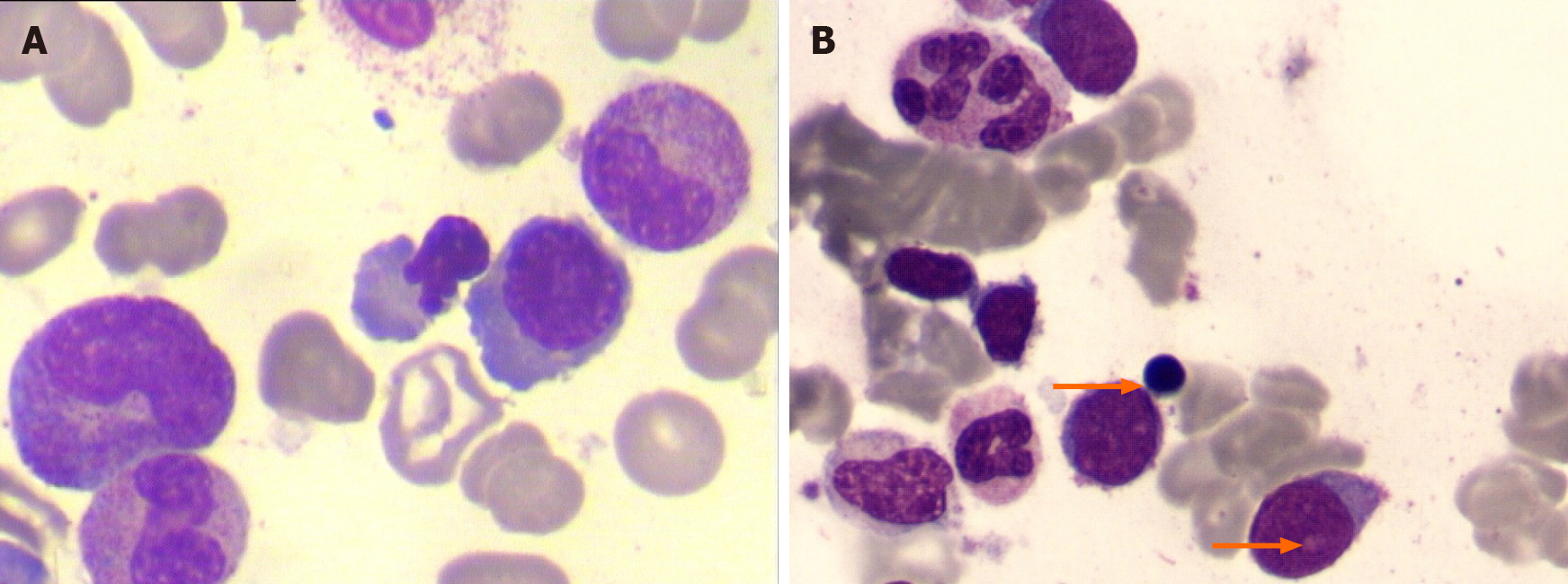
Figure 1 The first and third bone marrow examinations.
A: The first bone marrow examination showing single erythroid dysplasia without myeloblast; B: The third bone marrow examination revealing trilineage dysplasia and increased myeloblast (arrows).
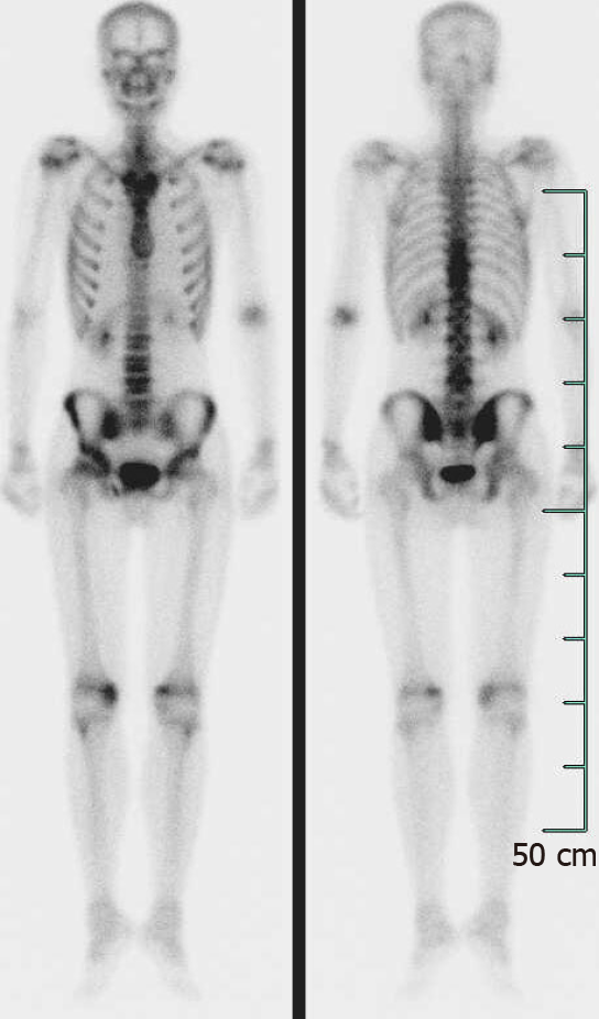
Figure 2 Tc-99m bone scintigraphy imaging.
It revealed increased radio-uptake in the T9-11, L4 vertebral, the right sacroiliac joint, and both knee joints.
Figure 3 X ray and computed tomography imaging.
A: X ray of the pelvis showing unbilateral asymmetric sacroiliitis, grade 3 on the right side and grade 2 on the left side; B: Computed tomography demonstrating narrowing space of sacroiliac joints, erosion, and subchondral sclerosis on the right iliac side.
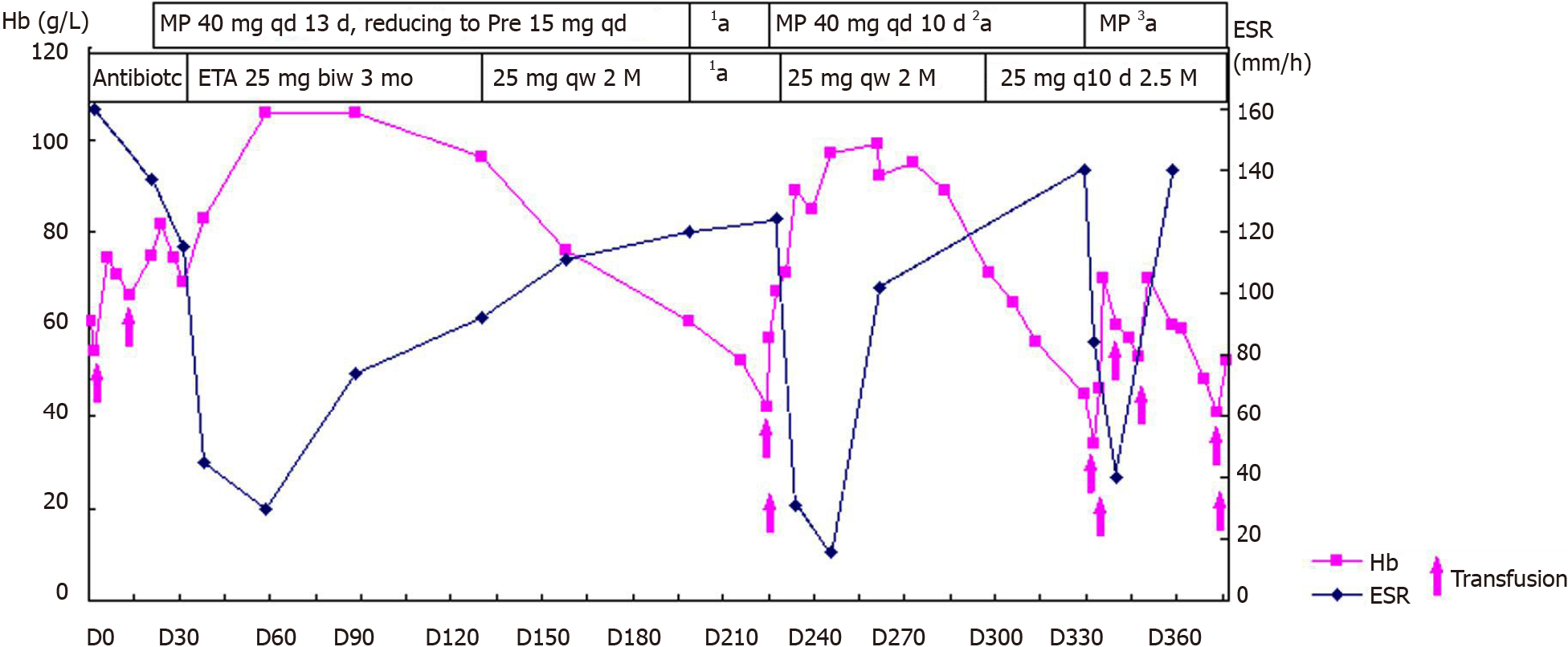
Figure 4 Curves of Hb and ESR levels before, during, and after treatment with etanercept, glucocorticoid, and transfusion.
MP: Methylprednisolone; Pre: prednisone; ETA: Etanercept; Hb: Hemoglobin; 1a: The patient self-discontinued etanercept and prednisone; 2a: Tapering to prednisone 25 mg daily; 3a: MP 40 mg daily for 10 d, be off for 24 d, then MP 20 mg daily for 8 d, tapering to prednisone 20 mg daily.
- Citation: Xu GH, Lin J, Chen WQ. Concurrent ankylosing spondylitis and myelodysplastic syndrome: A case report. World J Clin Cases 2022; 10(6): 1929-1936
- URL: https://www.wjgnet.com/2307-8960/full/v10/i6/1929.htm
- DOI: https://dx.doi.org/10.12998/wjcc.v10.i6.1929


