Copyright
©The Author(s) 2022.
World J Clin Cases. Feb 26, 2022; 10(6): 1922-1928
Published online Feb 26, 2022. doi: 10.12998/wjcc.v10.i6.1922
Published online Feb 26, 2022. doi: 10.12998/wjcc.v10.i6.1922
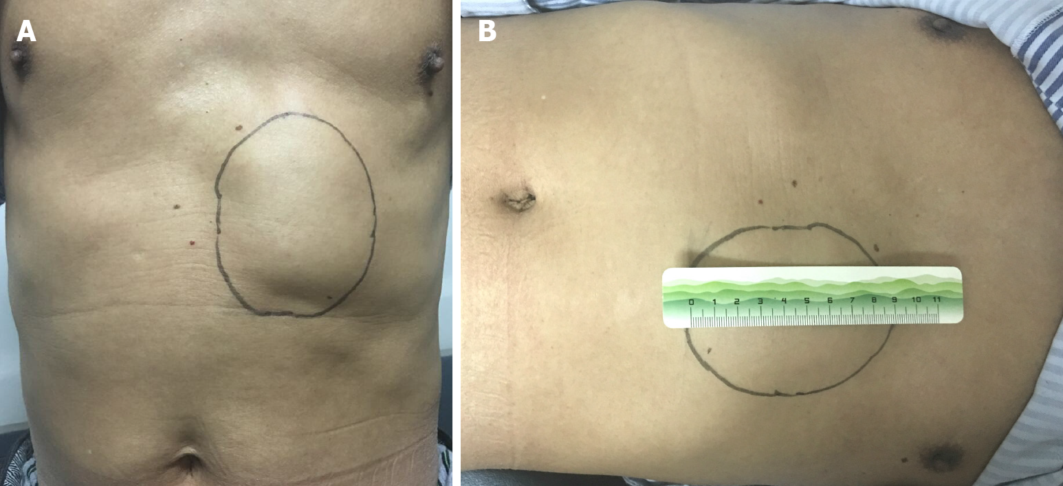
Figure 1 Physical examination of the mass.
A and B: Location and morphology of the abdominal wall mass in the preoperative patient.
Figure 2 Imaging findings of the mass.
A: The ultrasound showed that the tumor was a hypoechoic mass with an unclear boundary and an uneven internal echo (length 83 mm, and width 31 mm); B: Computed tomography (CT) scans showed inhomogeneous low contrast enhancement after injection of the contrast agent; C: The sagittal reconstruction of CT scans showed a solid space occupying the left anterior abdominal wall.
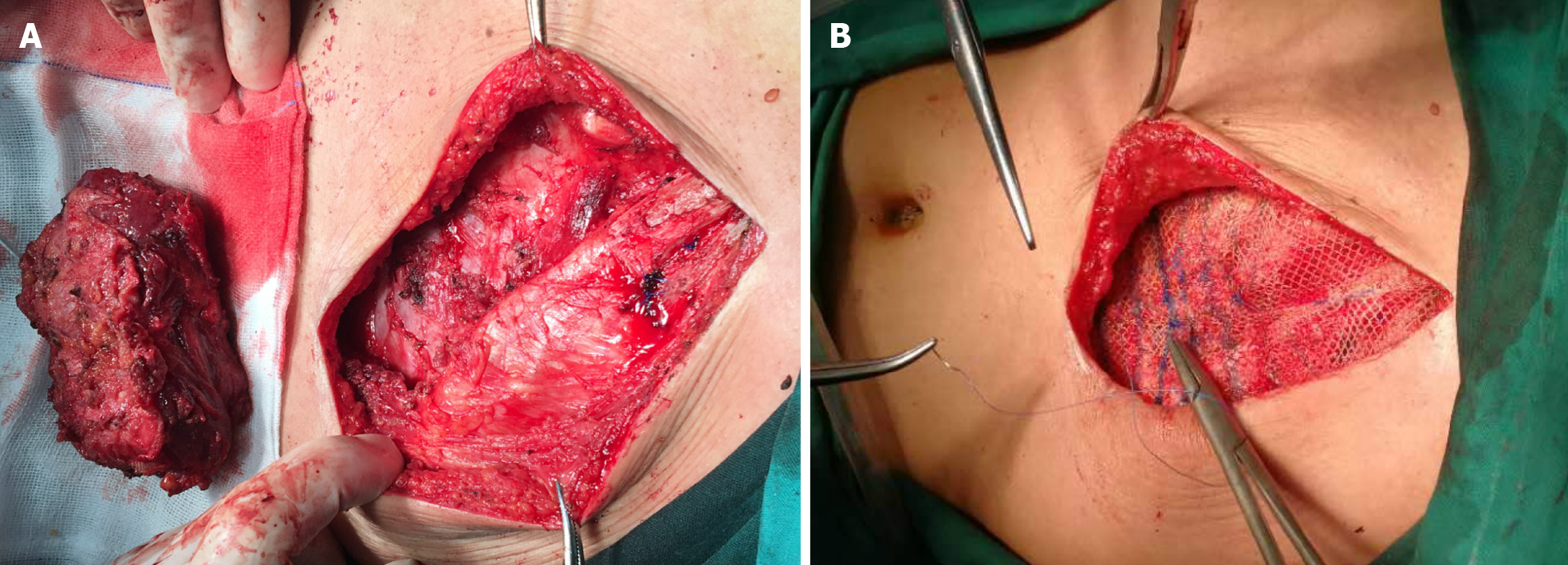
Figure 3 Resection of the mass (left position).
A: The removed mass and defect of the abdominal wall; B: The defect repaired with a patch.
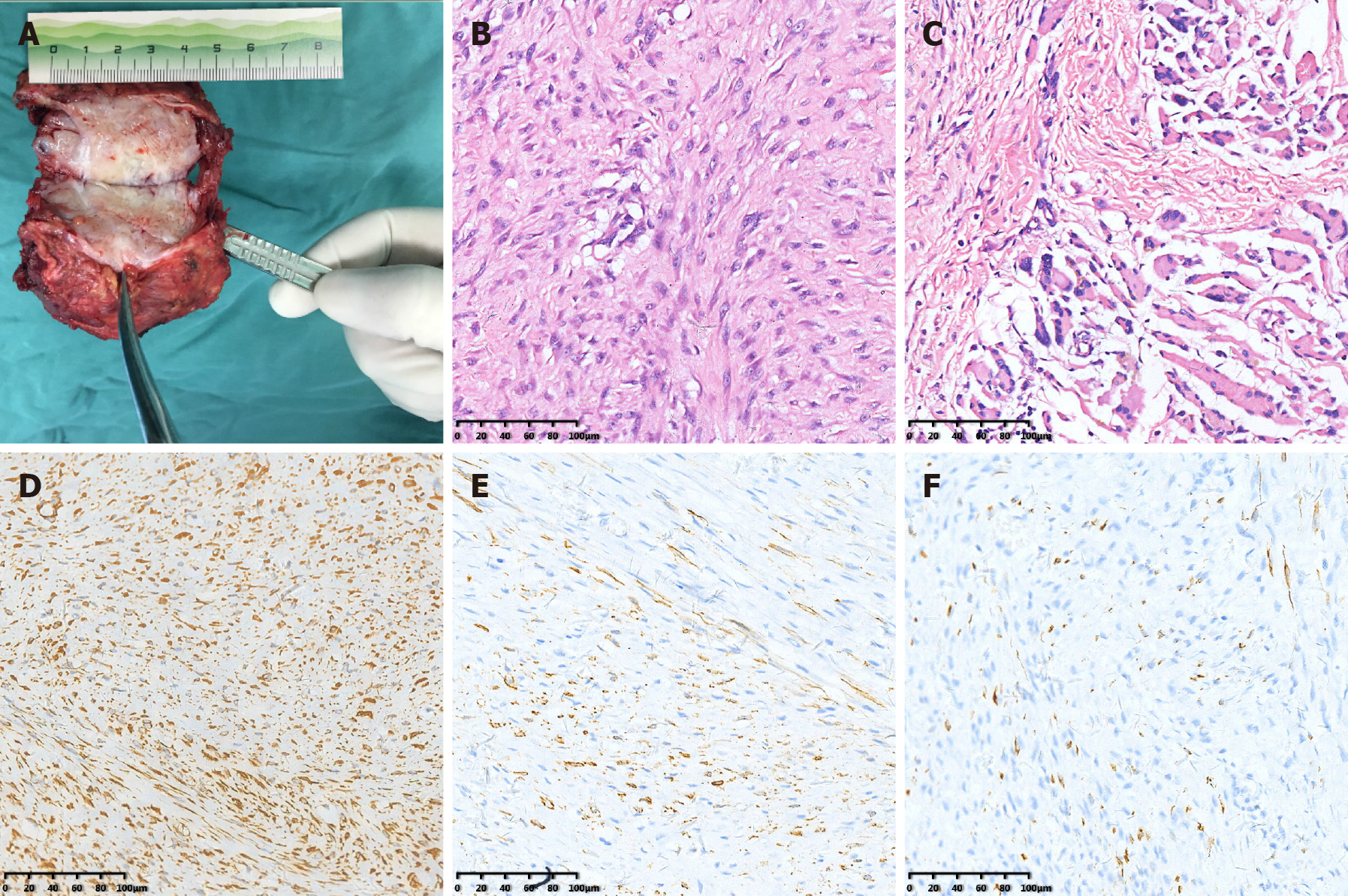
Figure 4 Patient pathology and immunohistochemistry.
A: A transverse section of the gross specimen showing fishy flesh; B: The tumor was composed of spindle cells and focally expressed ganglion-like cells (hematoxylin and eosin stained, 20 × magnification); C: The pathological examination showed that the lesion interspersed and grew between the rhabdomyo fibers, forming a checkerboard-like structure, not involving the rhabdomyo fibers themselves, while a large number of lymphocyte infiltrations could be seen locally (hematoxylin and eosin stained, 20 × magnification); D: Vimentin positive (20 × magnification); E: Smooth muscles actin positive (20 × magnification); F: Desmin positive (20 × magnification).
- Citation: Xing RW, Nie HQ, Zhou XF, Zhang FF, Mou YH. Left abdominal wall proliferative myositis resection and patch repair: A case report. World J Clin Cases 2022; 10(6): 1922-1928
- URL: https://www.wjgnet.com/2307-8960/full/v10/i6/1922.htm
- DOI: https://dx.doi.org/10.12998/wjcc.v10.i6.1922


