Copyright
©The Author(s) 2022.
World J Clin Cases. Feb 26, 2022; 10(6): 1914-1921
Published online Feb 26, 2022. doi: 10.12998/wjcc.v10.i6.1914
Published online Feb 26, 2022. doi: 10.12998/wjcc.v10.i6.1914
Figure 1 Cerebral corridor creator (Shineyard Medical, Shenzhen, Guangdong Province, China).
A: Balloon catheter set; B: Balloon catheter with inner needle details; C: Tubular retractor; D: Securing the tubular retractor by following the catheter route.
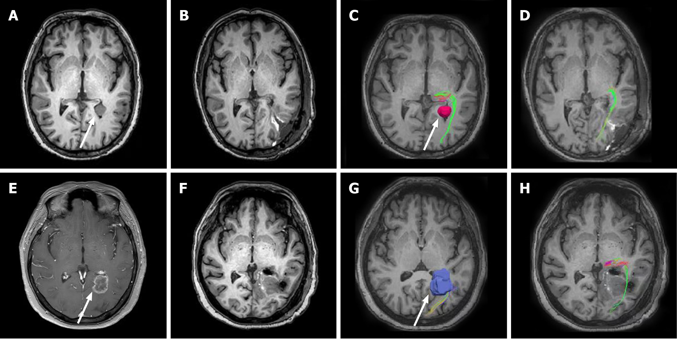
Figure 2 Comparative magnetic resonance imaging and diffusion tensor imaging from the axial plane.
A-D: Case 1: Preoperative T1-weighted image demonstrates a lesion in the left lateral ventricular trigone (A); Postoperative T1-weighted image demonstrates total tumor resection (B); Preoperative diffusion tensor imaging (DTI) shows the left optic radiation (OR) wrapped laterally around the tumor (C); Postoperative DTI shows the presence of intact OR adjacent to the tumor cavity was completely resected (D); E-H: Case 2: Preoperative T1-weighted image after Gadolinium contrast administration demonstrates a prominent ring-enhancing lesion in the left lateral ventricular trigone (E); Postoperative T1-weighted image demonstrates total tumor resection (F); Preoperative DTI reveals the left OR with subtle deformation due to tumor mass effect (G); Postoperative surgery: DTI shows the preserved left OR (H).
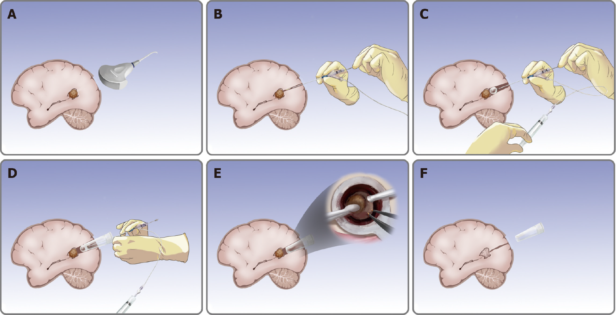
Figure 3 Removal of trigone ventricular tumors using cerebral corridor creator.
A: Check the tumor position; B: Puncture balloon catheter; C: Inflate and deflate the balloon; D: Secure the tubular retractor; E: Remove the tumor (details from the surgeon’s view); F: Withdraw the tubular retractor.
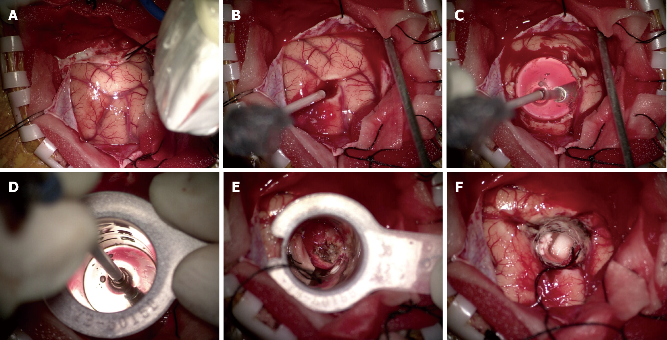
Figure 4 Case 1: Intra-operative photograph.
A: Intra-operative ultrasound relocated the tumor and the entry point; B: Puncture balloon catheter; C: Inflate the balloon; D: Secure the tubular retractor; E: Tumor is seen within the tubular retractor; F: Total resection of the tumor.
- Citation: Liu XW, Lu WR, Zhang TY, Hou XS, Fa ZQ, Zhang SZ. Cerebral corridor creator for resection of trigone ventricular tumors: Two case reports. World J Clin Cases 2022; 10(6): 1914-1921
- URL: https://www.wjgnet.com/2307-8960/full/v10/i6/1914.htm
- DOI: https://dx.doi.org/10.12998/wjcc.v10.i6.1914


