Copyright
©The Author(s) 2022.
World J Clin Cases. Feb 26, 2022; 10(6): 1903-1908
Published online Feb 26, 2022. doi: 10.12998/wjcc.v10.i6.1903
Published online Feb 26, 2022. doi: 10.12998/wjcc.v10.i6.1903
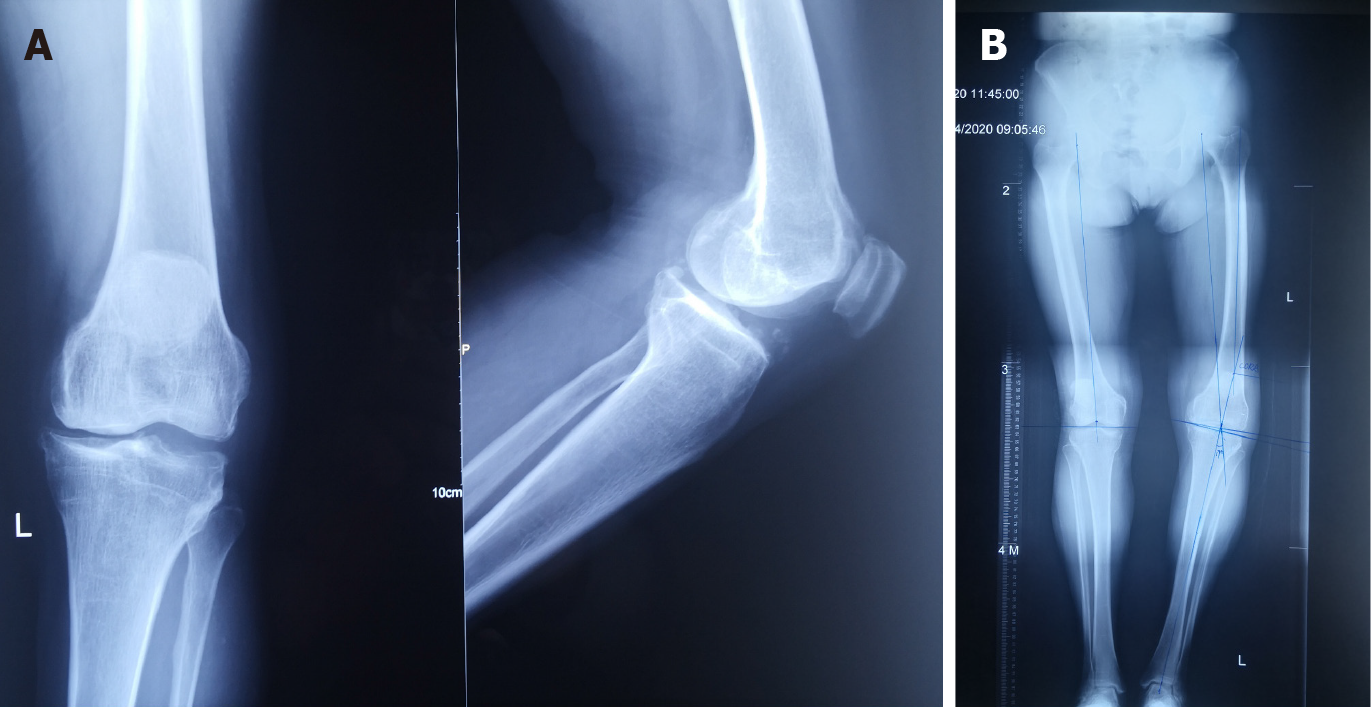
Figure 1 Preoperative imaging examination.
A: Preoperative anteroposterior and lateral radiographs of left knee; B: Using long-leg weight-bearing radiograph before surgery to measure the angle of lower limbs.
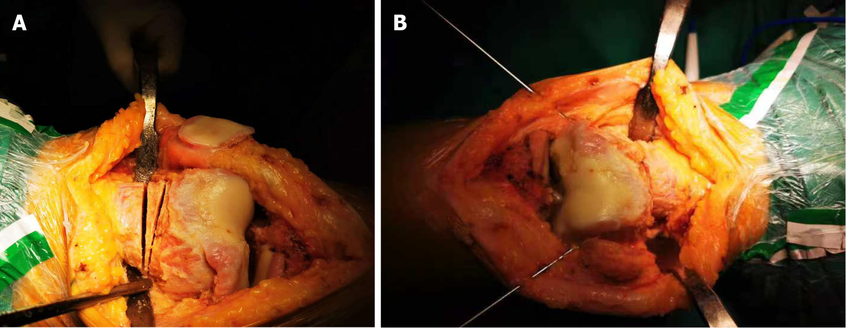
Figure 2 Intraoperative findings.
A: Intraoperative wedge osteotomy was performed on the supracondylar femur according to CORA principle; B: After reduction, Kirschner wires were fixed on both sides of the femoral osteotomy.
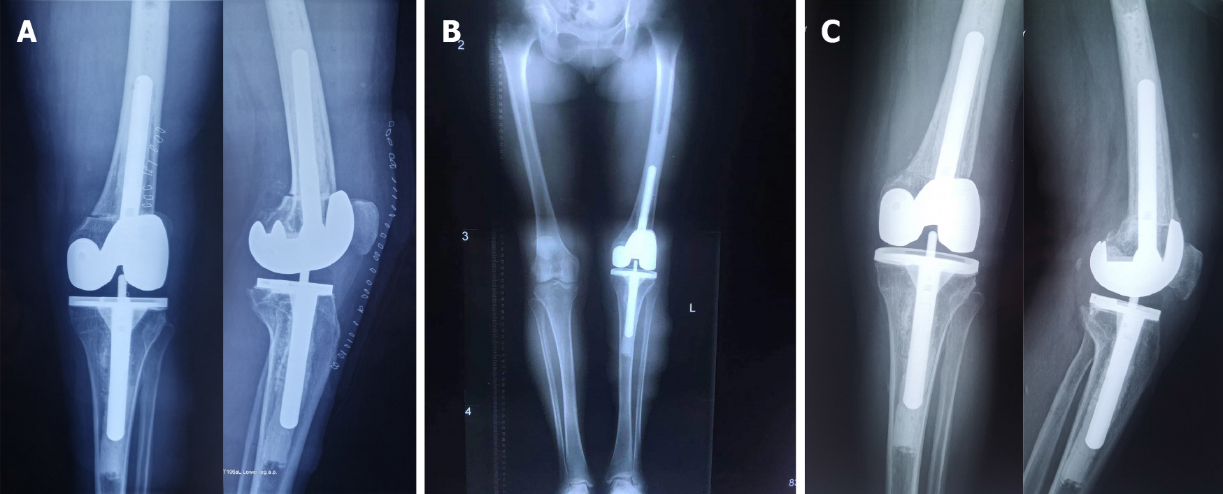
Figure 3 Postoperative imaging examination.
A: Postoperative anteroposterior and lateral radiographs of left knee; B: Long-leg weight-bearing radiograph after surgery; C: Anteroposterior and lateral radiographs of the left knee taken 8 mo after surgery.
- Citation: Xu SM, Li W, Zhang DB, Bi HY, Gu GS. Modified treatment of knee osteoarthritis complicated with femoral varus deformity: A case report. World J Clin Cases 2022; 10(6): 1903-1908
- URL: https://www.wjgnet.com/2307-8960/full/v10/i6/1903.htm
- DOI: https://dx.doi.org/10.12998/wjcc.v10.i6.1903


