Copyright
©The Author(s) 2022.
World J Clin Cases. Feb 6, 2022; 10(4): 1432-1440
Published online Feb 6, 2022. doi: 10.12998/wjcc.v10.i4.1432
Published online Feb 6, 2022. doi: 10.12998/wjcc.v10.i4.1432
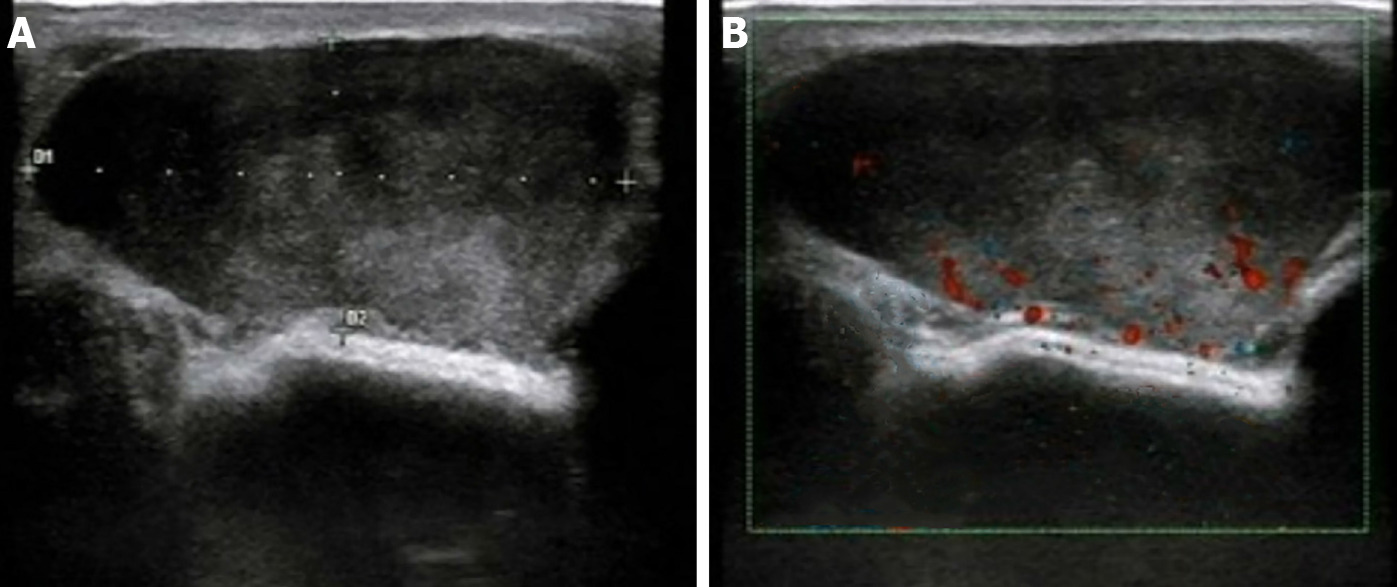
Figure 1 Ultrasound manifestations of inflammatory myofibroblastic tumor.
A: An oval, hypoechoic mass with clear borders of approximately 4.2 cm × 1.8 cm in size was identified at the 9 o’clock position in the left breast. The internal echo was heterogeneous, with scattered small fleck echo and slightly enhanced rear echo; B: Color Doppler flow imaging indicated limited blood flow signal within the hypoechoic mass.
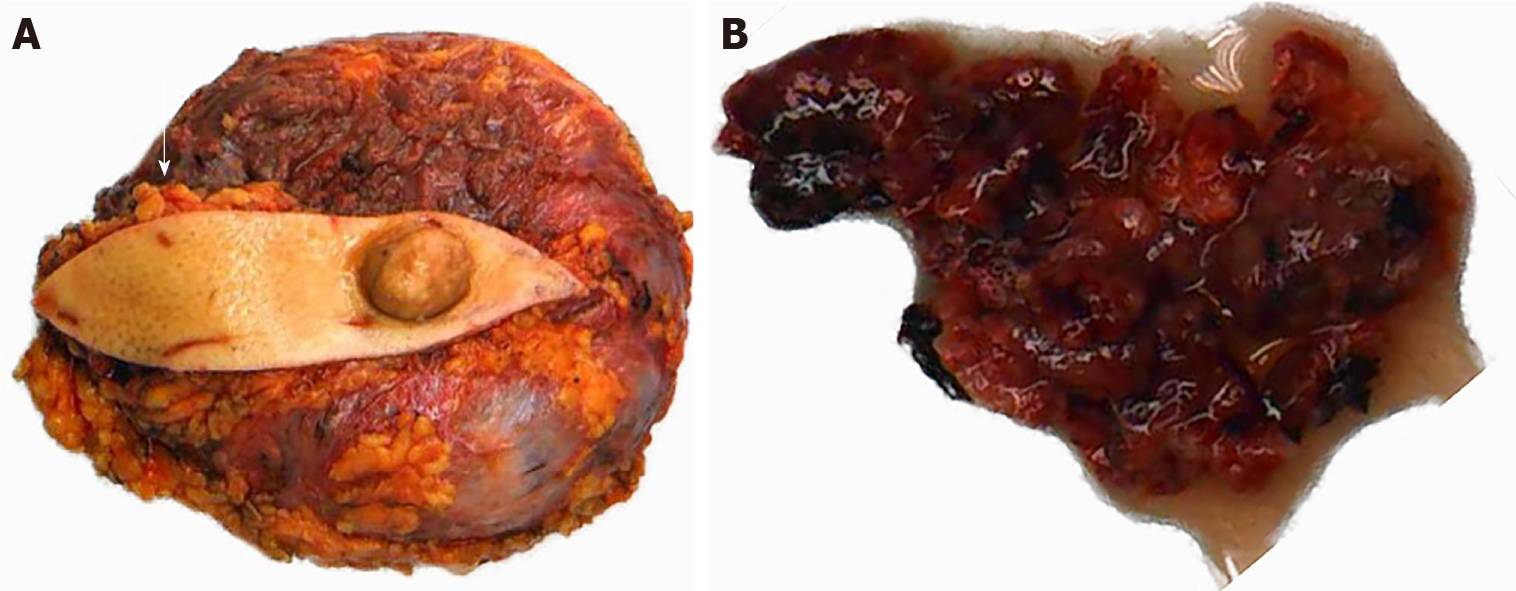
Figure 2 Tumor and prosthesis in the left breast, with fusiform skin flap and nipple.
A: A prosthesis was observed in the breast tissue, with a subcutaneous nodule identified 3.5 cm away from the nipple (arrow); B: The left breast mass was grayish red and soft, with partial tissue deletion.
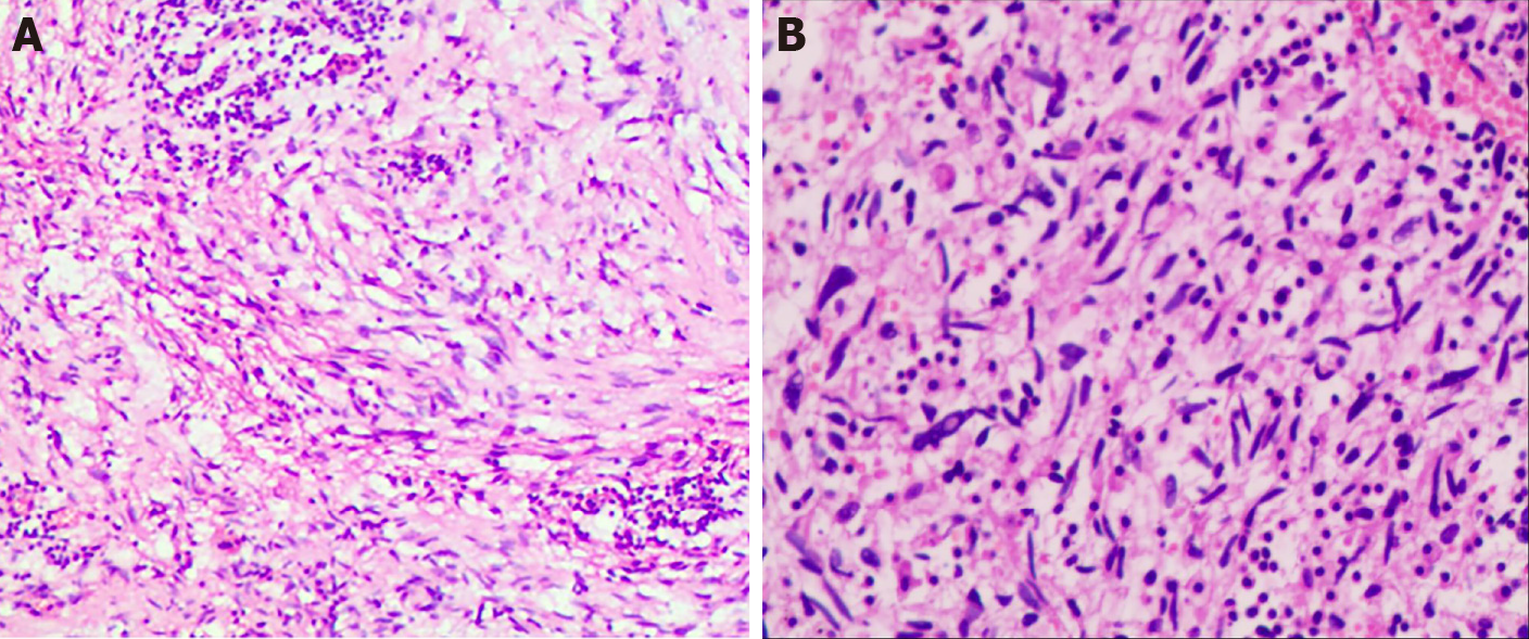
Figure 3 Histopathological findings of inflammatory myofibroblastic tumor.
A: Hematoxylin-eosin (HE) staining (magnification, × 100) revealed that the spindle cells were diffusely proliferated, irregularly arranged, and scattered in the nucleus, with mildly atypical cells, mitosis, interstitial vascular proliferation, and dilation and congestion with hemorrhage; B: HE staining (magnification, × 200) revealed that a large number of neutrophils and lymphocytes and plasma cell infiltration were observed.
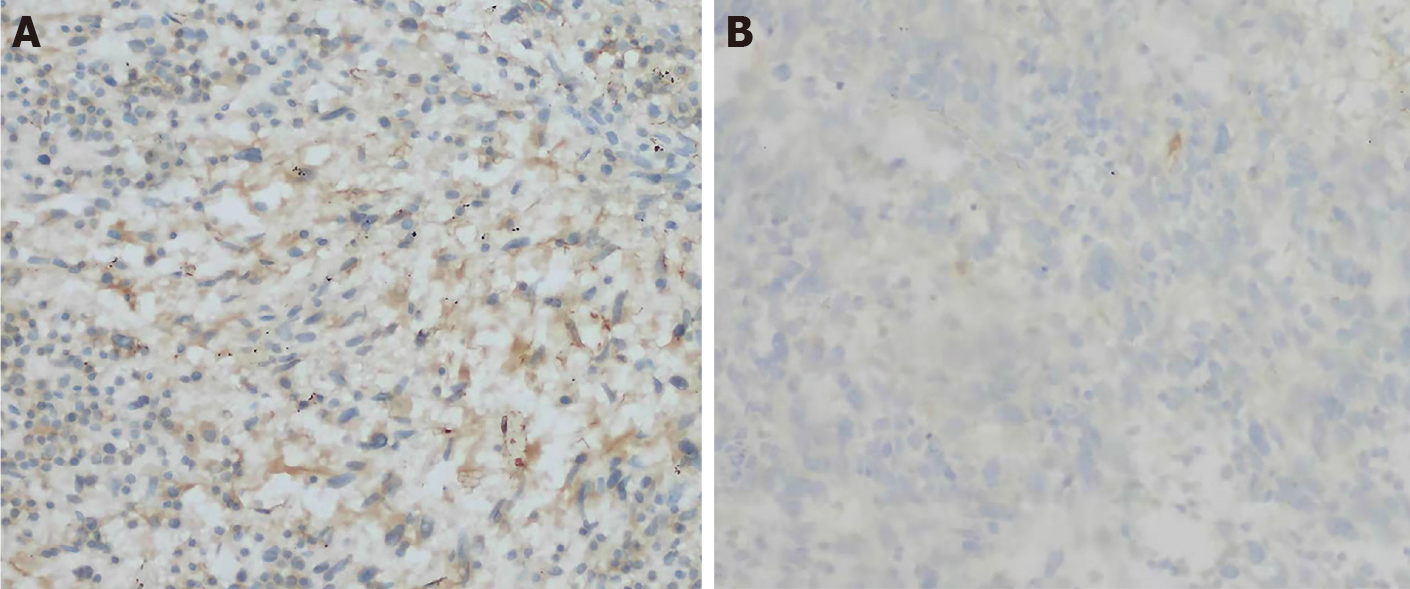
Figure 4 Immunohistochemical staining.
A: The cells were partially positive for smooth muscle actin (magnification, ×200); B: Negative for anaplastic lymphoma kinase (magnification, ×200).
- Citation: Zhou P, Chen YH, Lu JH, Jin CC, Xu XH, Gong XH. Inflammatory myofibroblastic tumor after breast prosthesis: A case report and literature review. World J Clin Cases 2022; 10(4): 1432-1440
- URL: https://www.wjgnet.com/2307-8960/full/v10/i4/1432.htm
- DOI: https://dx.doi.org/10.12998/wjcc.v10.i4.1432


