Copyright
©The Author(s) 2022.
World J Clin Cases. Feb 6, 2022; 10(4): 1410-1416
Published online Feb 6, 2022. doi: 10.12998/wjcc.v10.i4.1410
Published online Feb 6, 2022. doi: 10.12998/wjcc.v10.i4.1410
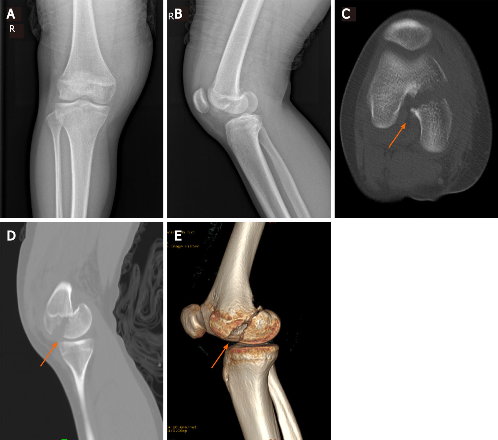
Figure 1 Image data before operative treatment showing a distal femoral medial condylar fracture.
A and B: Anteroposterior and lateral radiographs images before operative treatment; C and D: Computed tomography images before operative treatment (arrow); E: Computed tomography three dimensional reconstruction images before operative treatment (arrow).
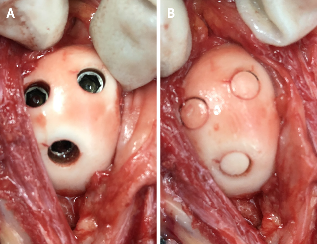
Figure 2 Intraoperative images.
A: Image after screws fixation and countersunk; B: Image after osteochondral plugs covering the screw heads.
Figure 3 Images of the affected joint, 6 mo postoperatively.
A: Complete extension of the right knee joint; B: Complete flexion of the right knee joint; C: Posteromedial approach.
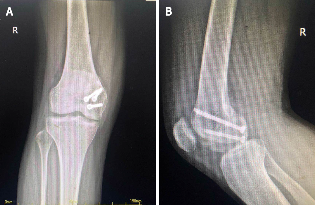
Figure 4 Image data of 6 mo after operative treatment showing a healed fracture and osteochondral plug.
A: Anteroposterior radiographs images after operative treatment; B: Lateral radiographs images after operative treatment.
- Citation: Jiang ZX, Wang P, Ye SX, Xie XP, Wang CX, Wang Y. Hoffa’s fracture in an adolescent treated with an innovative surgical procedure: A case report. World J Clin Cases 2022; 10(4): 1410-1416
- URL: https://www.wjgnet.com/2307-8960/full/v10/i4/1410.htm
- DOI: https://dx.doi.org/10.12998/wjcc.v10.i4.1410


