Copyright
©The Author(s) 2022.
World J Clin Cases. Feb 6, 2022; 10(4): 1401-1409
Published online Feb 6, 2022. doi: 10.12998/wjcc.v10.i4.1401
Published online Feb 6, 2022. doi: 10.12998/wjcc.v10.i4.1401
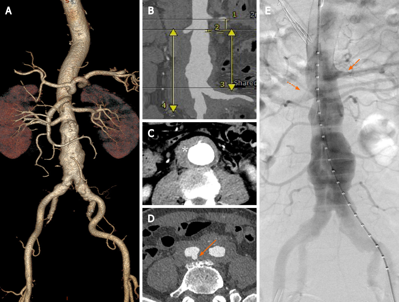
Figure 1 Preoperative imaging.
A: 3D reconstruction of the aorta; B: Proximally healthy, 20-mm-long landing zone between renal arteries; C: Perivascular hematoma (asterisk); D: Penetrating aortic ulcers near the bifurcation of the right iliac artery (solid arrow); E: Angiography consistent with preoperative computed tomography angiography.
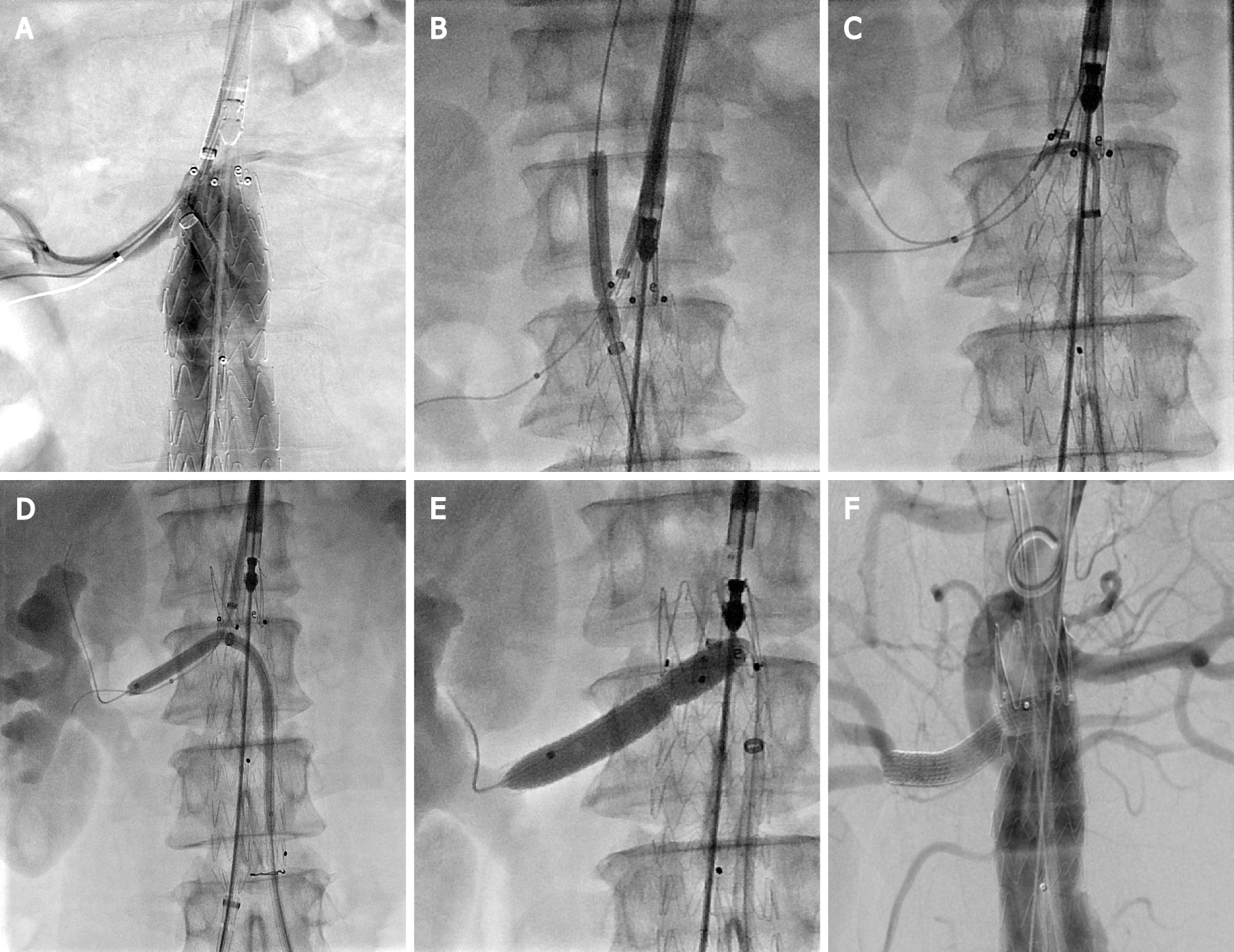
Figure 2 Steps for fenestrations.
A: The tip of the steerable sheath was adjusted to align with the ostium of the right renal artery (RRA); B: Progressive enlargement of the fenestration was performed using balloons; C: A trimmed pigtail catheter was used to guide the guidewire to the RRA; D: The bare stent region was released with a balloon anchored at the RRA; E: The covered bridging stent was deployed; F: Angiography demonstrated that the RRA was patent, with no endoleakage.
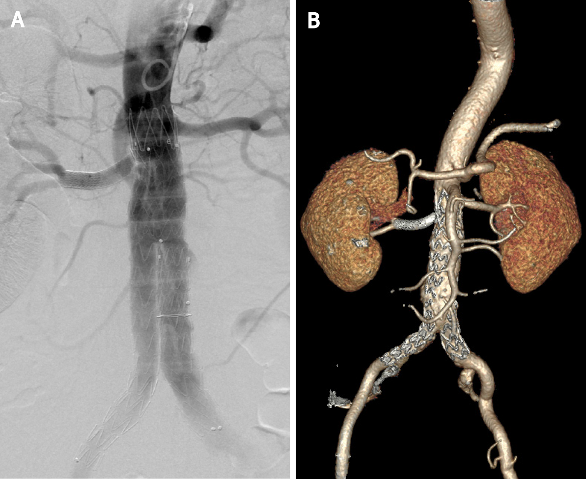
Figure 3 Postoperative imaging.
A: Final angiogram; B: Computed tomography reconstruction at 1 year demonstrated graft patency.
- Citation: Wang ZW, Qiao ZT, Li MX, Bai HL, Liu YF, Bai T. Antegrade in situ laser fenestration of aortic stent graft during endovascular aortic repair: A case report. World J Clin Cases 2022; 10(4): 1401-1409
- URL: https://www.wjgnet.com/2307-8960/full/v10/i4/1401.htm
- DOI: https://dx.doi.org/10.12998/wjcc.v10.i4.1401


