Copyright
©The Author(s) 2022.
World J Clin Cases. Feb 6, 2022; 10(4): 1394-1400
Published online Feb 6, 2022. doi: 10.12998/wjcc.v10.i4.1394
Published online Feb 6, 2022. doi: 10.12998/wjcc.v10.i4.1394
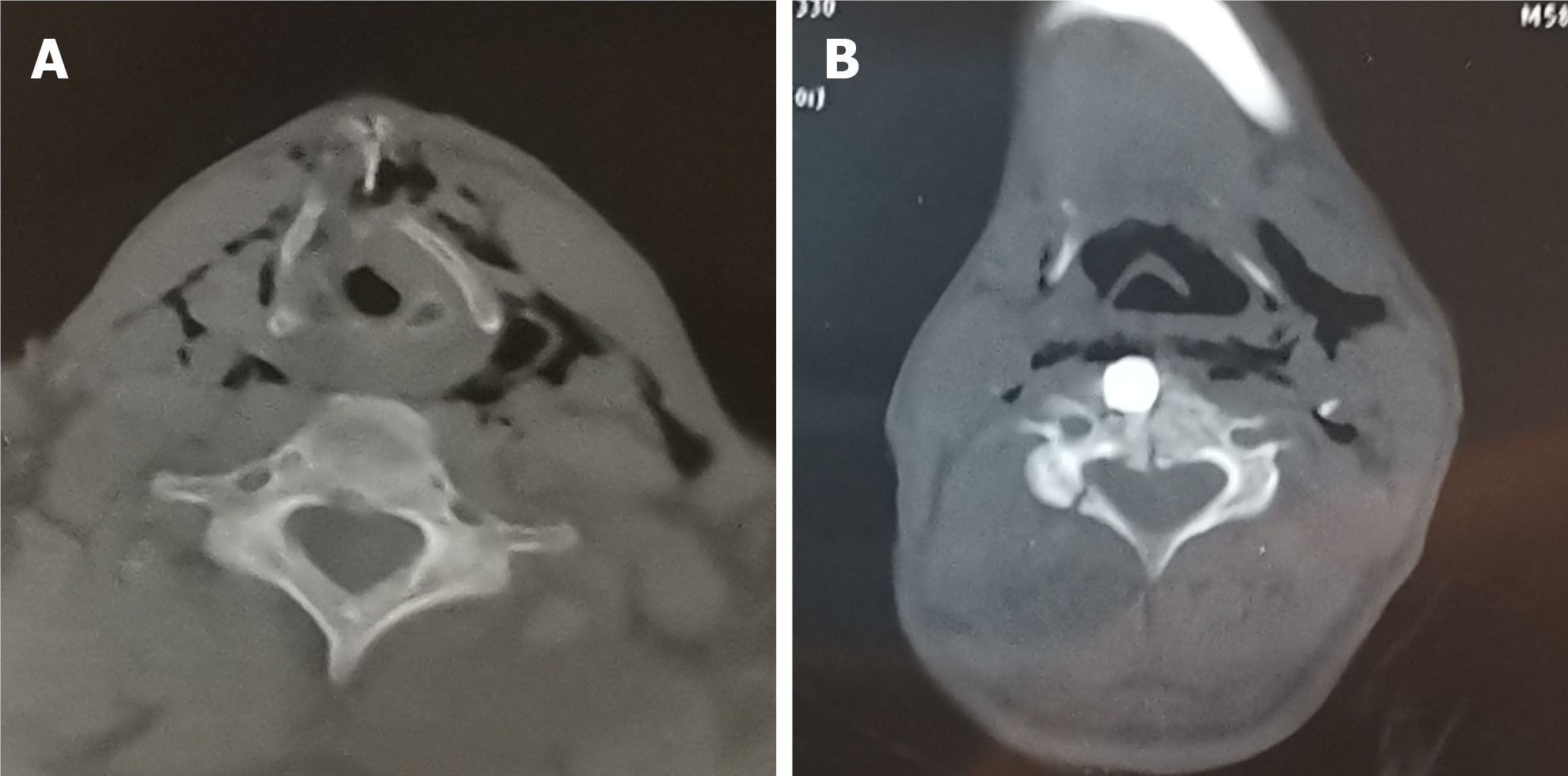
Figure 1 Axial computed tomography scan of the neck.
A: Laryngeal injury; B: Metal fragment.
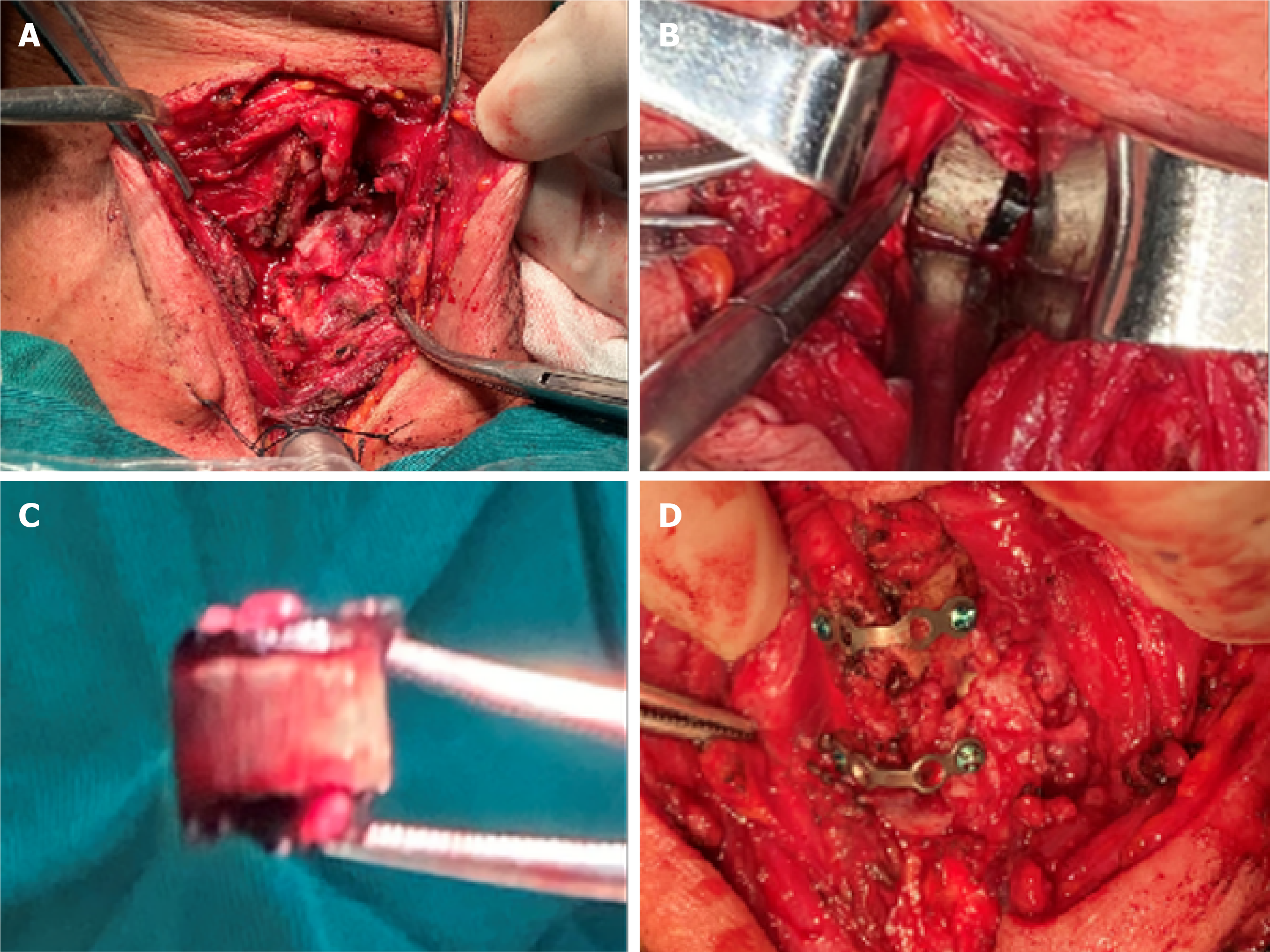
Figure 2 Intra-operative images.
A: Laryngeal injury; B: Fragment lodged in the C4 vertebra; C: Fragment was removed; D: Miniplate fixation.
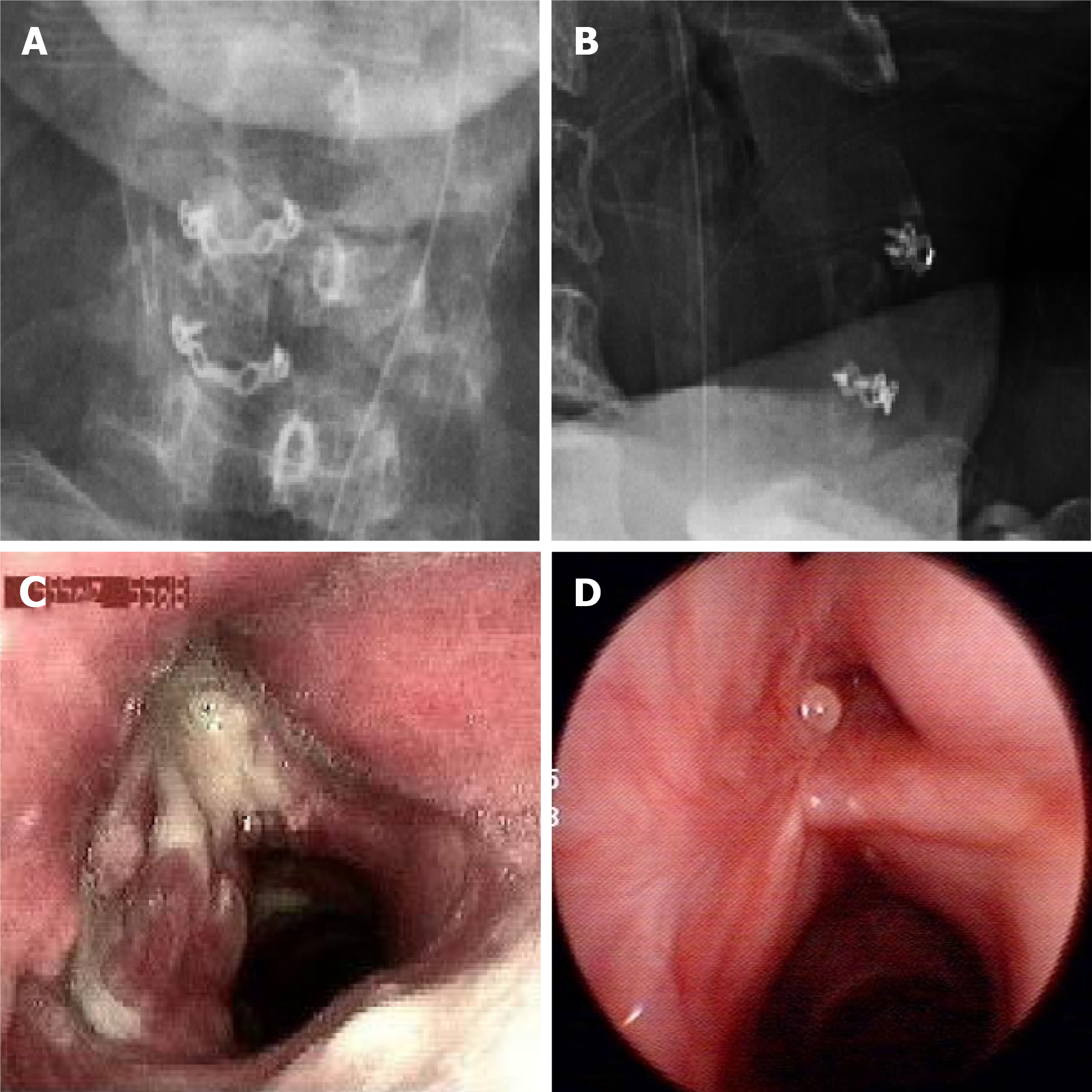
Figure 3 Post-operative findings.
A: Antero-posterior radiograph, demonstrating good position of the miniplates; B: Lateral radiograph, demonstrating good position of the miniplates; C: Fibrolaryngoscopy on the 14th day after surgery, demonstrating the condition of the endolarynx; D: Dynamic laryngoscopy 6 mo after surgery, demonstrating good recovery of the larynx.
- Citation: Qiu ZH, Zeng J, Zuo Q, Liu ZQ. External penetrating laryngeal trauma caused by a metal fragment: A Case Report. World J Clin Cases 2022; 10(4): 1394-1400
- URL: https://www.wjgnet.com/2307-8960/full/v10/i4/1394.htm
- DOI: https://dx.doi.org/10.12998/wjcc.v10.i4.1394


