Copyright
©The Author(s) 2022.
World J Clin Cases. Feb 6, 2022; 10(4): 1373-1380
Published online Feb 6, 2022. doi: 10.12998/wjcc.v10.i4.1373
Published online Feb 6, 2022. doi: 10.12998/wjcc.v10.i4.1373
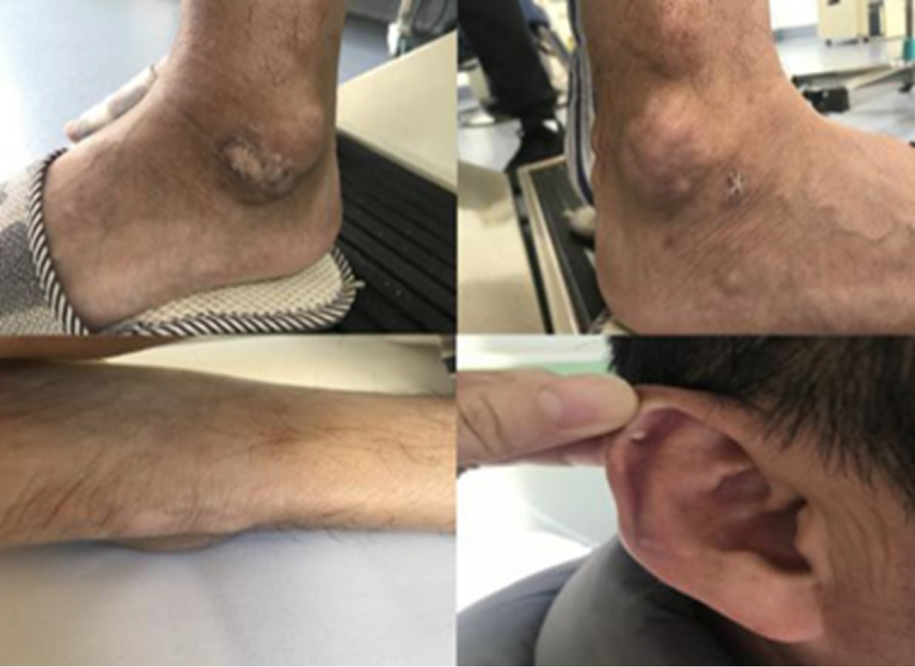
Figure 1 Multiple gouty tophi.
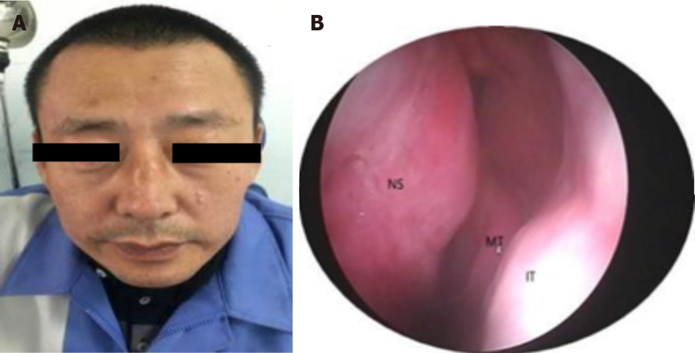
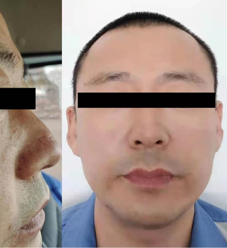
Figure 2 Preoperative physical examination.
A: Preoperative frontal view; B: Nasal endoscope, nasal septum: obvious hypertrophy and swelling of the left upper mucosa of the nasal septum, middle turbinate, inferior turbinate. NS: Nasal septum; MT: Middle turbinate; IT: Inferior turbinate.
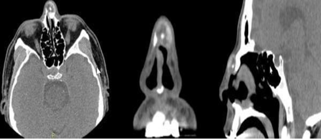
Figure 3 The head and neck computed tomography scan displayed a mottled radiopaque mass crossed over the nasal ridge.
The boundary was clear, the size approximately 1.7 cm × 1.3 cm, the plain scan computed tomography (CT) value approximately 22-96 HU, the enhanced scan CT values approximately 25-104 HU and 14-109 HU, and the left nasal bone locally discontinuous.
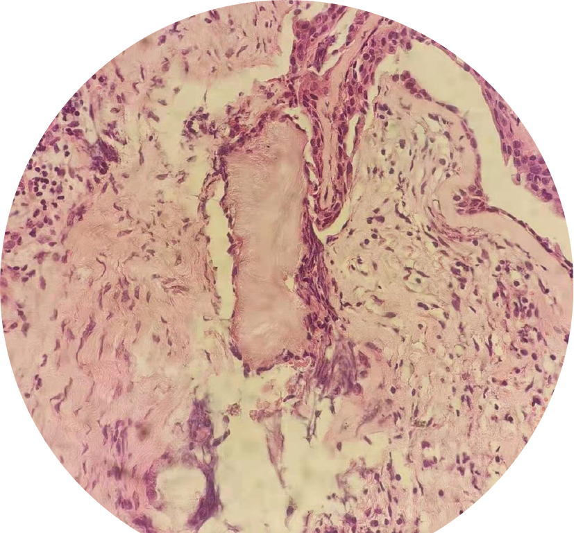
Figure 4 Postoperative histopathological examination.
Figure 5 One year after operation, there was no deformity of nasal bridge and no recurrence of gouty tophus.
- Citation: Song Y, Kang ZW, Liu Y. Multiple gouty tophi in the head and neck with normal serum uric acid: A case report and review of literatures. World J Clin Cases 2022; 10(4): 1373-1380
- URL: https://www.wjgnet.com/2307-8960/full/v10/i4/1373.htm
- DOI: https://dx.doi.org/10.12998/wjcc.v10.i4.1373


