Copyright
©The Author(s) 2022.
World J Clin Cases. Feb 6, 2022; 10(4): 1357-1365
Published online Feb 6, 2022. doi: 10.12998/wjcc.v10.i4.1357
Published online Feb 6, 2022. doi: 10.12998/wjcc.v10.i4.1357
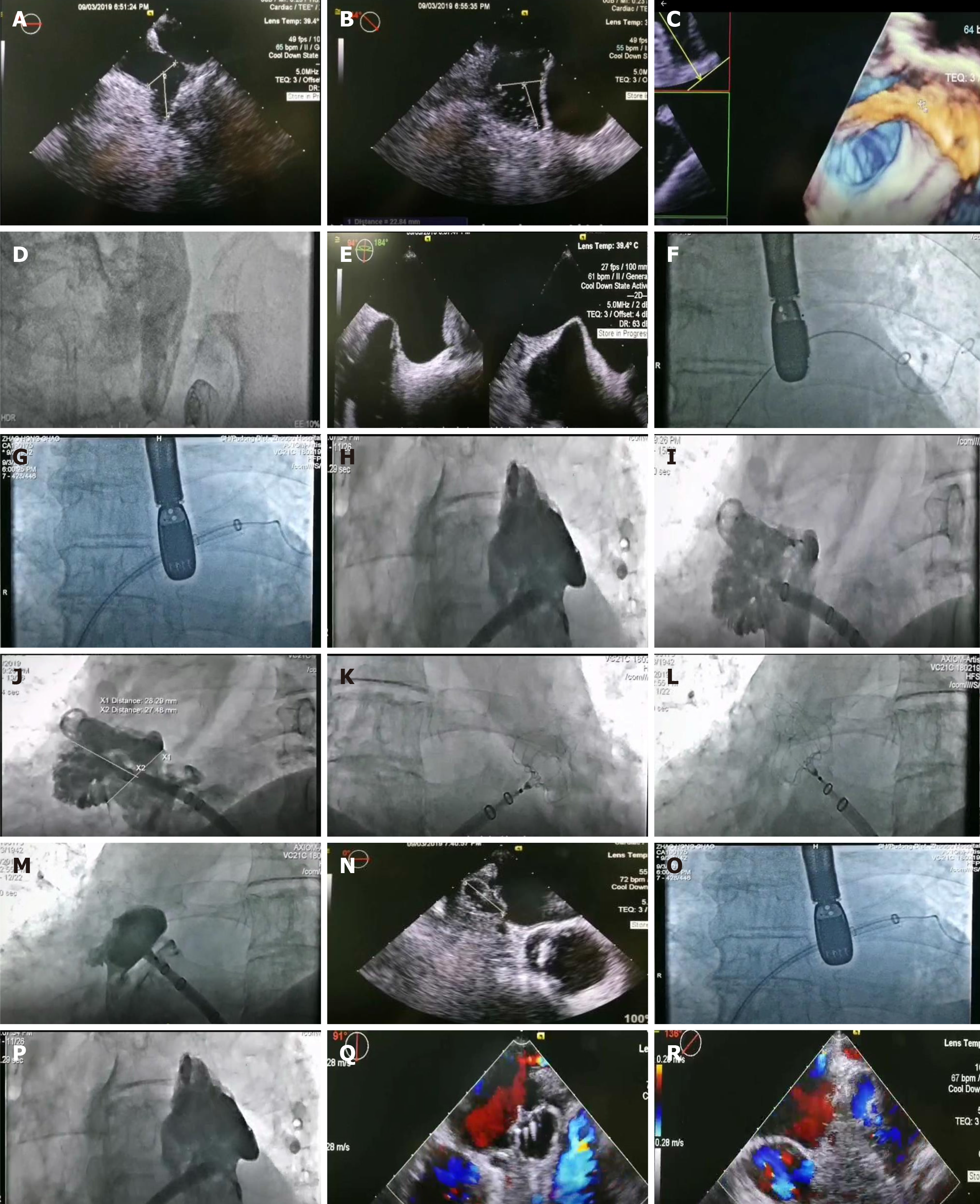
Figure 1 Details of the surgical procedure.
A: Transesophageal echocardiography; B: Dextrocardia (45°); C: Morphology of left atrial appendage; D: Left femoral vein puncture; E: Atrial septum puncture; F: Superior pulmonary vein of guide wire; G: Sheath follow up; H: Auricle not fully exposed; I: Right anterior oblique cardiography; J: Occluder selection; K: Implantation of sealing umbrella; L: Pulling and plugging the closure umbrella; M: Release the closure umbrella; N: Multi angle compression ratio (24%-27%); O: No residual shunt (0°); P: 1.5mm residual shunt (45°); Q: No residual shunt (90°); R: No residual shunt (135°).
- Citation: Tian B, Ma C, Su JW, Luo J, Sun HX, Su J, Ning ZP. Left atrial appendage occlusion in a mirror-image dextrocardia: A case report and review of literature. World J Clin Cases 2022; 10(4): 1357-1365
- URL: https://www.wjgnet.com/2307-8960/full/v10/i4/1357.htm
- DOI: https://dx.doi.org/10.12998/wjcc.v10.i4.1357


