Copyright
©The Author(s) 2022.
World J Clin Cases. Feb 6, 2022; 10(4): 1341-1348
Published online Feb 6, 2022. doi: 10.12998/wjcc.v10.i4.1341
Published online Feb 6, 2022. doi: 10.12998/wjcc.v10.i4.1341
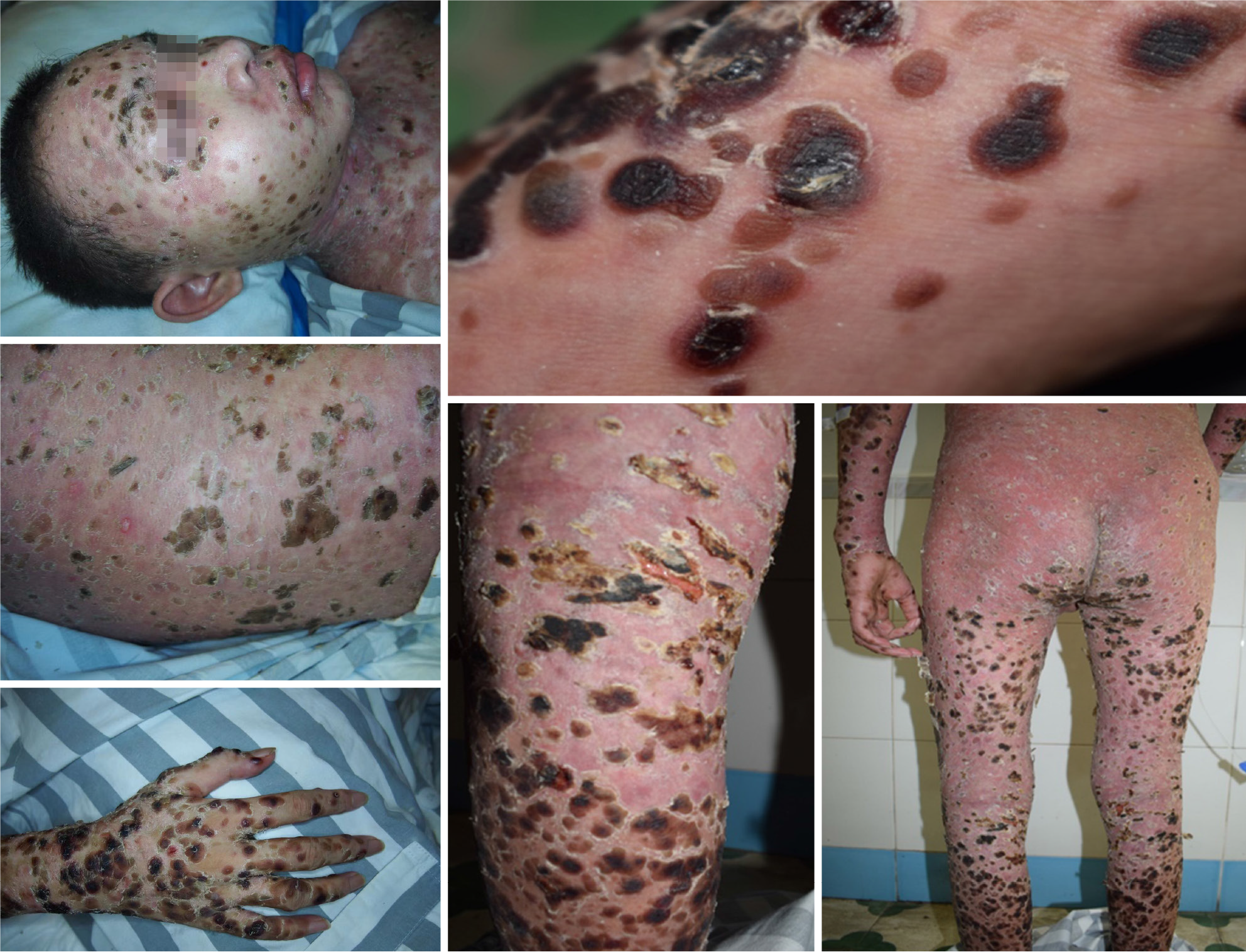
Figure 1 Photographs of skin lesions before treatment.
Scattered blood blisters were observed throughout the body, with the most severe blood blisters on the trunk, consistent with the distribution of skin transverse striae.
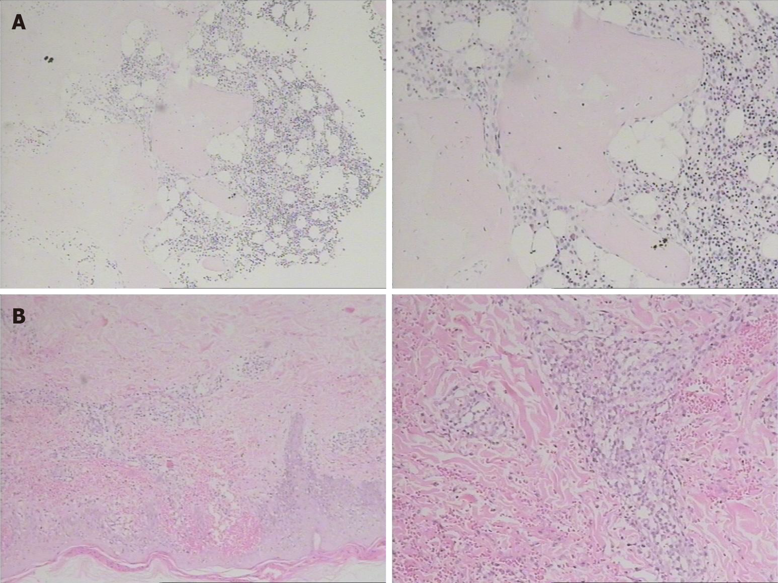
Figure 2 Bone marrow and skin biopsies.
A: Bone marrow biopsy. Slight microscopic bone marrow hyperplasia, cell volume accounted for 40%, tertiary hematopoietic cells were present, granulocyte/erythrocyte ratio was slightly increased, and cell morphology was normal; B: Skin biopsy of left thigh. There was hyperkeratosis of the skin epidermis, hemorrhage in the papilla of the dermis, and local or diffuse small lymphocyte infiltration in both the epidermis and subcutaneously.
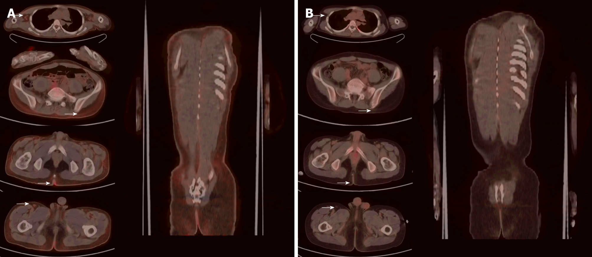
Figure 3 The patient underwent positron emission tomography/computed tomography before treatment and reexamination after 4 cycles of treatment.
A: Positron emission tomography/computed tomography (PET/CT) before treatment. The figure showed mild systemic skin swelling and diffuse mild increase in glucose metabolism, especially in the local skin of both armpits, right upper quadrant, and posterior coccyx. Multiple small lymph nodes of different sizes with increased glucose metabolism were observed in both armpits and groins; B: PET/CT after treatment. The figure showed no clear structural or glucose metabolism abnormalities.
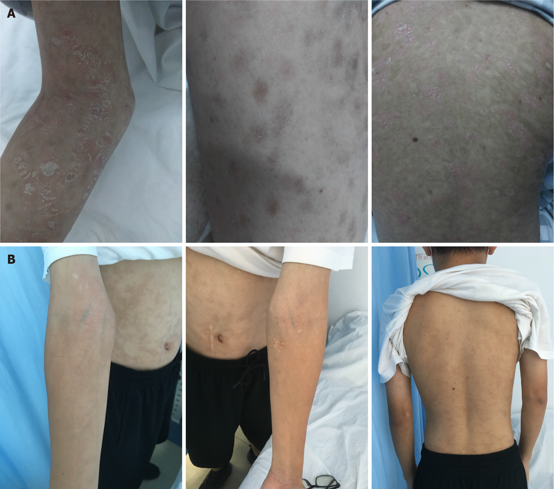
Figure 4 Follow-up photographs of the patient.
A: Photographs of the patient after completion of 4 treatment cycles. Generalized scattered blood blisters disappeared, and residual scattered skin pigmentation was observed; B: Photographs of the last follow-up. No abnormal lesions were observed.
- Citation: He ZD, Yang HY, Zhou SS, Wang M, Mo QL, Huang FX, Peng ZG. Chidamide combined with traditional chemotherapy for primary cutaneous aggressive epidermotropic CD8+ cytotoxic T-cell lymphoma: A case report. World J Clin Cases 2022; 10(4): 1341-1348
- URL: https://www.wjgnet.com/2307-8960/full/v10/i4/1341.htm
- DOI: https://dx.doi.org/10.12998/wjcc.v10.i4.1341


