Copyright
©The Author(s) 2022.
World J Clin Cases. Feb 6, 2022; 10(4): 1278-1285
Published online Feb 6, 2022. doi: 10.12998/wjcc.v10.i4.1278
Published online Feb 6, 2022. doi: 10.12998/wjcc.v10.i4.1278
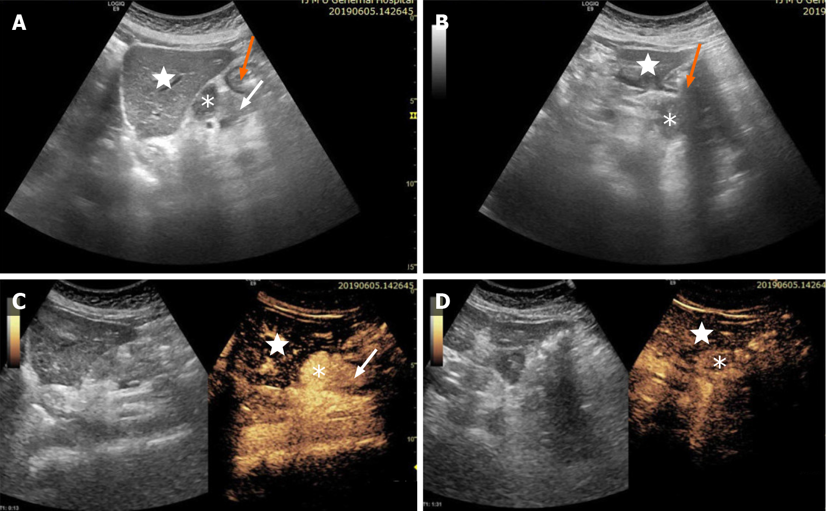
Figure 1 Patient underwent contrast-enhanced ultrasound examination.
Conventional and contrast-enhanced ultrasound of the hypoechoic mass (stellate) between the body of the pancreas (white arrow), left lobe of the liver (white star) and stomach (orange arrow), which has clear boundaries, irregular shape, uneven echoes, and no obvious blood flow signals. The hypoechoic mass showed homogeneous hyperenhancement in the arterial phase, and slightly high enhancement in the venous phase, and measured approximately 56 mm × 37 mm × 25 mm.
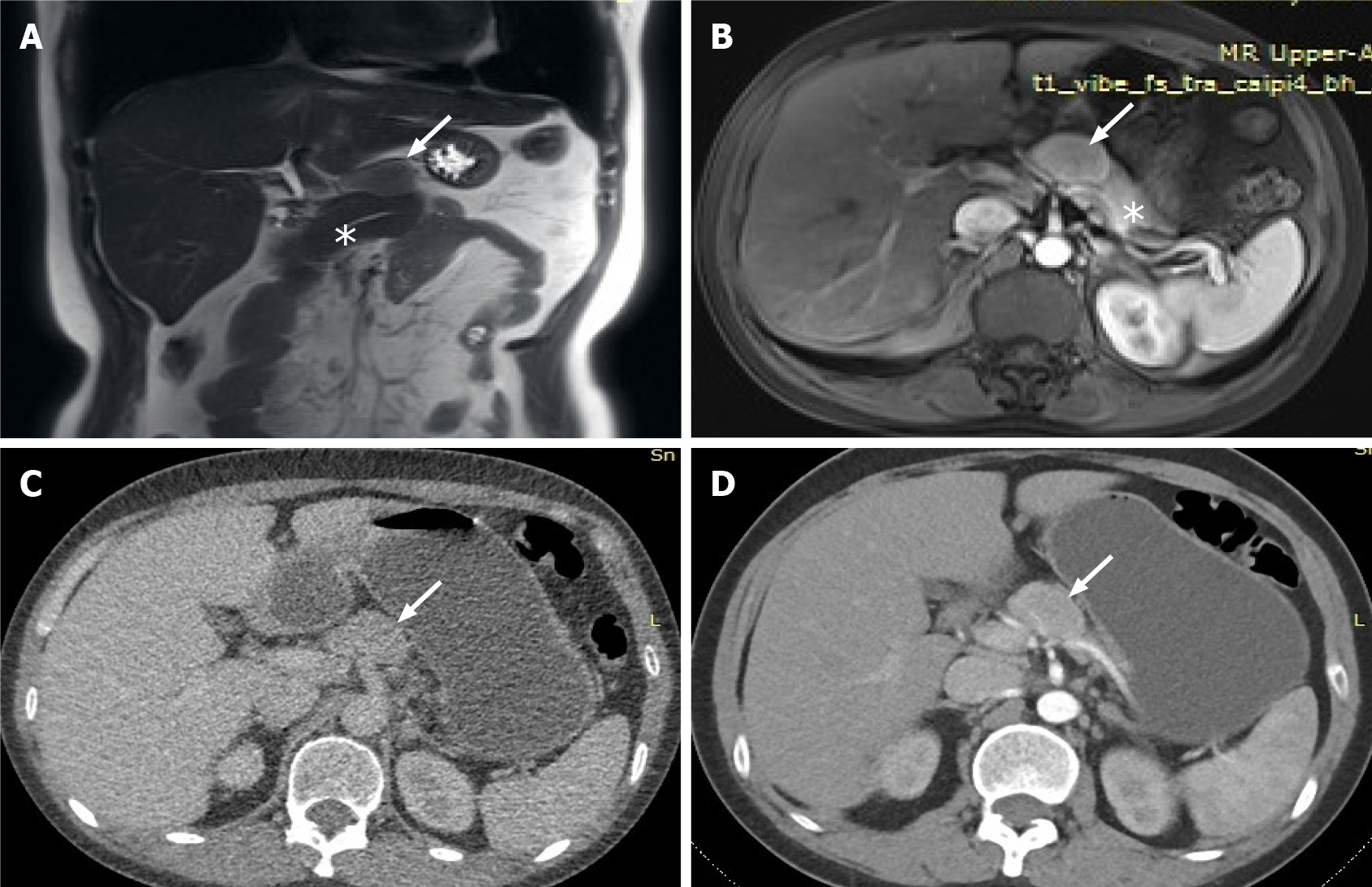
Figure 2 The lesion was further evaluated by computed tomography and magnetic resonance imaging.
A: Magnetic resonance imaging (MRI) showed a lesion of isointensity (white arrow) located above the pancreas body (stellate) which was clearly separated from the normal pancreas; B: Contrast-enhanced -MRI revealed that the edge was enhanced in the arterial phase, and the degree of internal enhancement in each phase was lower than that of the pancreatic parenchyma; C: Computed tomography (CT) showed a nodule of mixed density (white arrow) located above the pancreatic body, which was convex and shallowly lobulated, measuring approximately 37 mm 25 mm in maximum dimensions; D: Contrast-enhanced CT showed uneven progressive enhancement, and the degree of enhancement in each stage was lower than that of pancreatic parenchyma.
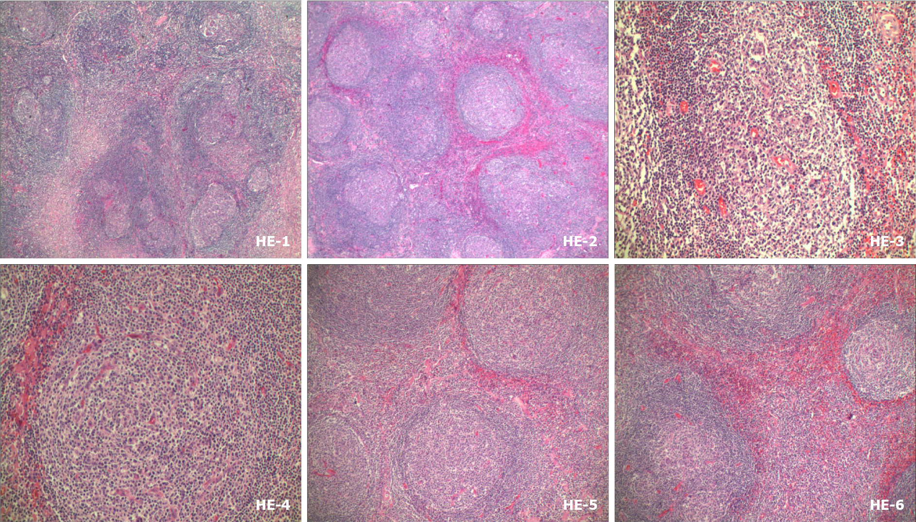
Figure 3
Hematoxylin and eosin staining revealed lymphoid tissue proliferation, with lymphoid follicular hyperplasia, mantle hyperplasia, interfollicular plasma cell hyperplasia, and local vascular hyperplasia.
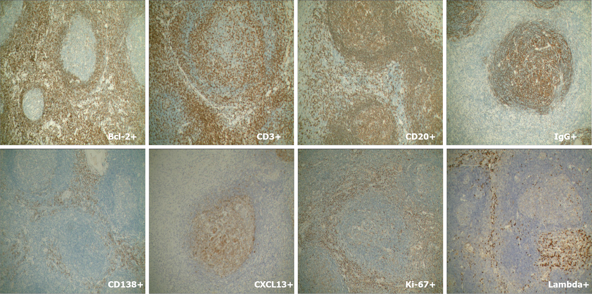
Figure 4 Plasma cells showed strong immunostaining for CD138 and IgG.
IgG4 was occasionally positive and the expression rate was < 40%. No restrictive expression of kappa and lambda was observed B and T cells were positive for CD20 and CD30. CD21 and CD23 showed the FDC network. Germinal center cells were positive for Bcl-2, and weakly positive for CD10 and Bcl-6. Cxc113 and cyclin D11 were positive in a few scattered cells. Ki-67 was highly expressed in germinal centers.
- Citation: Zhai HY, Zhu XY, Zhou GM, Zhu L, Guo DD, Zhang H. Unicentric Castleman disease was misdiagnosed as pancreatic mass: A case report. World J Clin Cases 2022; 10(4): 1278-1285
- URL: https://www.wjgnet.com/2307-8960/full/v10/i4/1278.htm
- DOI: https://dx.doi.org/10.12998/wjcc.v10.i4.1278


