Copyright
©The Author(s) 2022.
World J Clin Cases. Feb 6, 2022; 10(4): 1255-1262
Published online Feb 6, 2022. doi: 10.12998/wjcc.v10.i4.1255
Published online Feb 6, 2022. doi: 10.12998/wjcc.v10.i4.1255
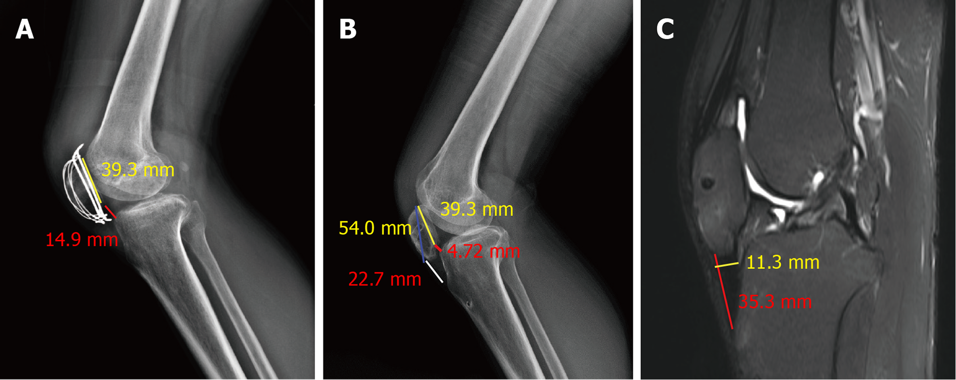
Figure 1 Preoperative lateral radiograph of the left knee and measurement of the patellar height.
A: Before the second operation, the fracture healed, and the Caton–Deschamps index was 0.38; B: Before the third operation. The ratio of the red dotted line and yellow dotted line indicates that the Caton–Deschamps index was 0.12, and the ratio of the white dotted line to the blue dotted line indicated that the Insall–Salvati index was 0.42; C: The contracted patellar tendon is approximately 35.0 mm in length and 11.3 mm in thickness.
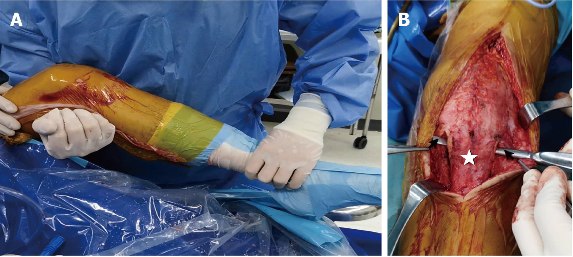
Figure 2 Arthroscopic release measurements were followed by range of motion (ROM) measurements and open surgery.
A: ROM of the knee joint after arthroscopic release was slightly improved compared with the preoperative ROM; B: The approach was made from 5 cm above the superior pole of the patella to the tibial tubercle. A periosteal elevator (black arrow) was used to mobilize the contracted patellar tendon (white star) and identify its medial and lateral margins.
Figure 3 Schematic diagram of the lengthening method.
A: The patellar tendon was longitudinally split along the midline while preserving the medial patellar insertion and the lateral tibial insertion of the patellar tendon (red line); B: Ultrabraid sutures were used to overlap the two tendon-free ends in a whipstitch fashion (blue line); C: The reconstructed patellar tendon was further strengthened using an iliotibial band graft (yellow area), which was fixed with two suture anchors (black circles), and suture cerclage was used to decrease tension (light blue line).
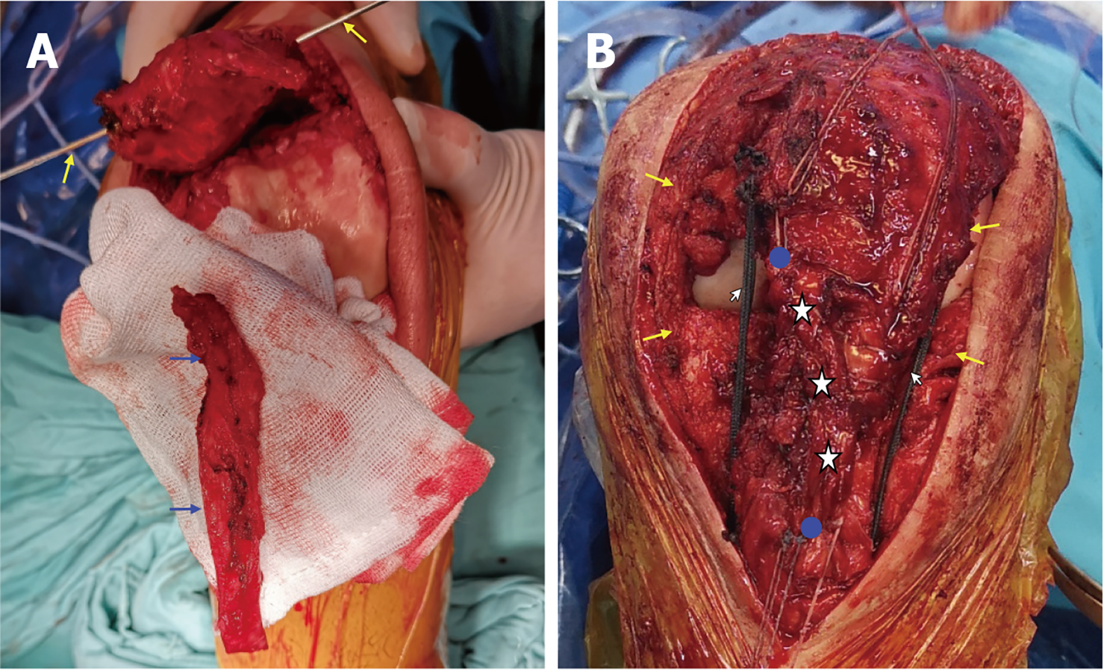
Figure 4 Intraoperative view of patellar tendon lengthening.
A: The Kirschner wire was used to drill a bone tunnel of the patella (thin yellow arrow), and the iliotibial band (ITB) graft was harvested to strengthen the reconstructed patellar tendon (blue arrow); B: Intraoperative view after patellar tendon reconstruction and ITB enhancement. Medial and lateral scar tissue surrounding the patellar tendon were removed (yellow arrow). The ITB graft (white star) was fixed with suture anchors (blue circle). Three MB66 polyester sutures were used as cerclage to decrease tension (white arrow).
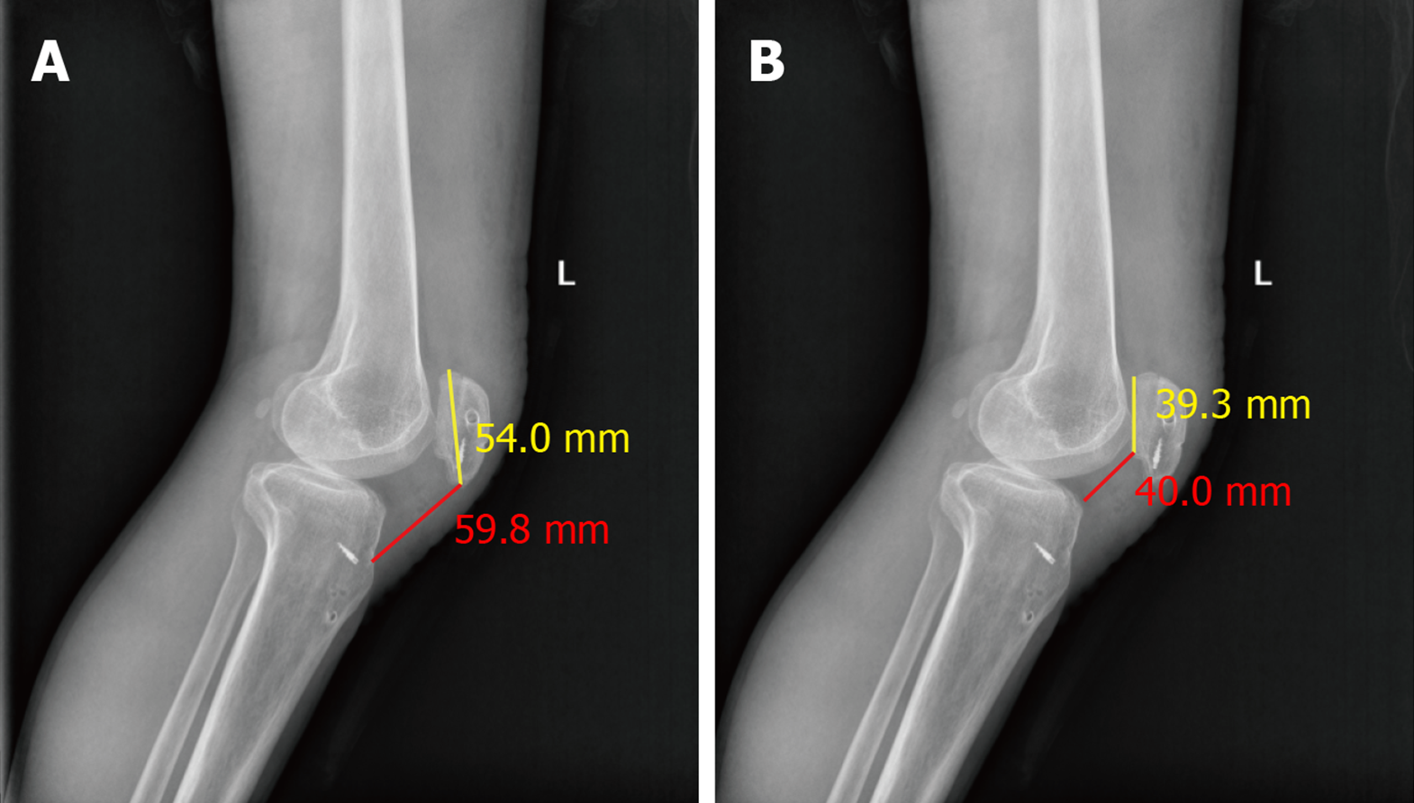
Figure 5 Radiographic image of the left knee postoperatively.
A: The Insall–Salvati index is 1.1; B: The Caton–Deschamps index is 1.0.
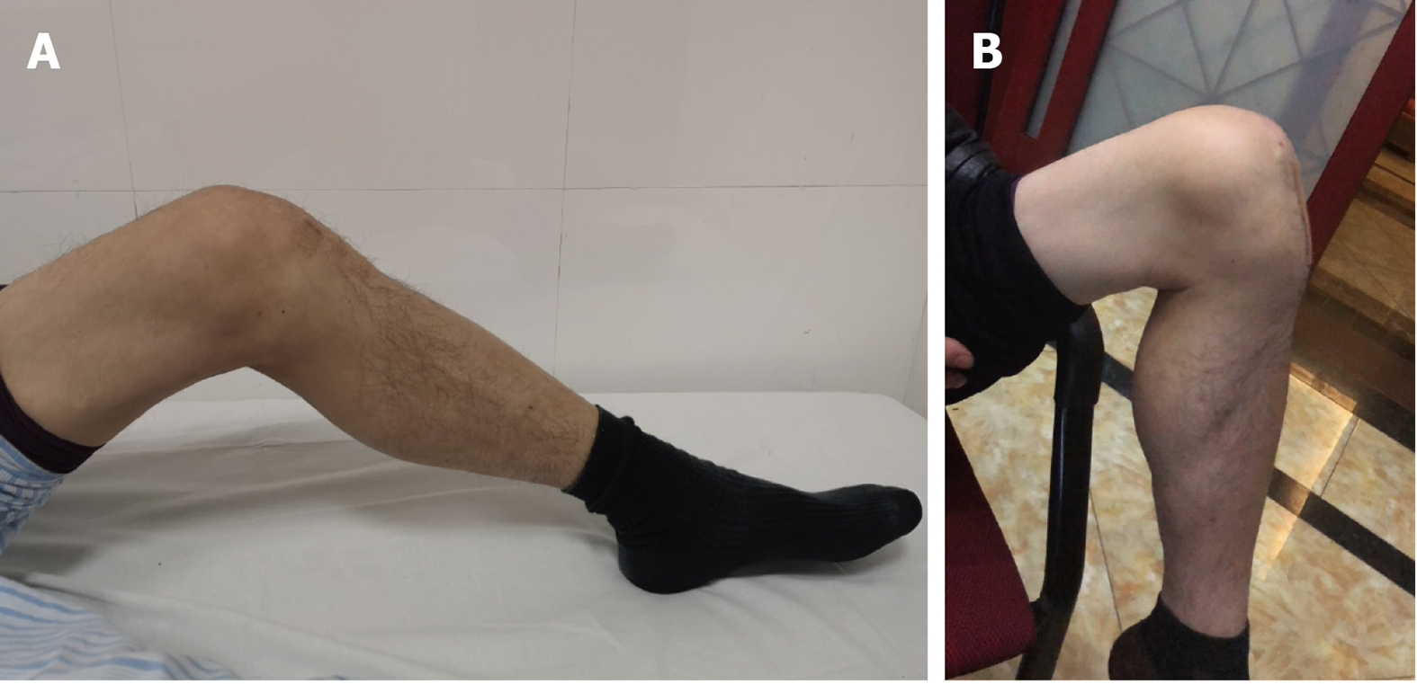
Figure 6 Comparison of the knee range of motion.
A: Preoperative range of motion (ROM) is approximately 0°-45°; B: The ROM improved to 0°–120° at the last follow-up.
- Citation: Tang DZ, Liu Q, Pan JK, Chen YM, Zhu WH. Autogenous iliotibial band enhancement combined with tendon lengthening plasty to treat patella baja: A case report. World J Clin Cases 2022; 10(4): 1255-1262
- URL: https://www.wjgnet.com/2307-8960/full/v10/i4/1255.htm
- DOI: https://dx.doi.org/10.12998/wjcc.v10.i4.1255


