Copyright
©The Author(s) 2022.
World J Clin Cases. Dec 26, 2022; 10(36): 13426-13434
Published online Dec 26, 2022. doi: 10.12998/wjcc.v10.i36.13426
Published online Dec 26, 2022. doi: 10.12998/wjcc.v10.i36.13426
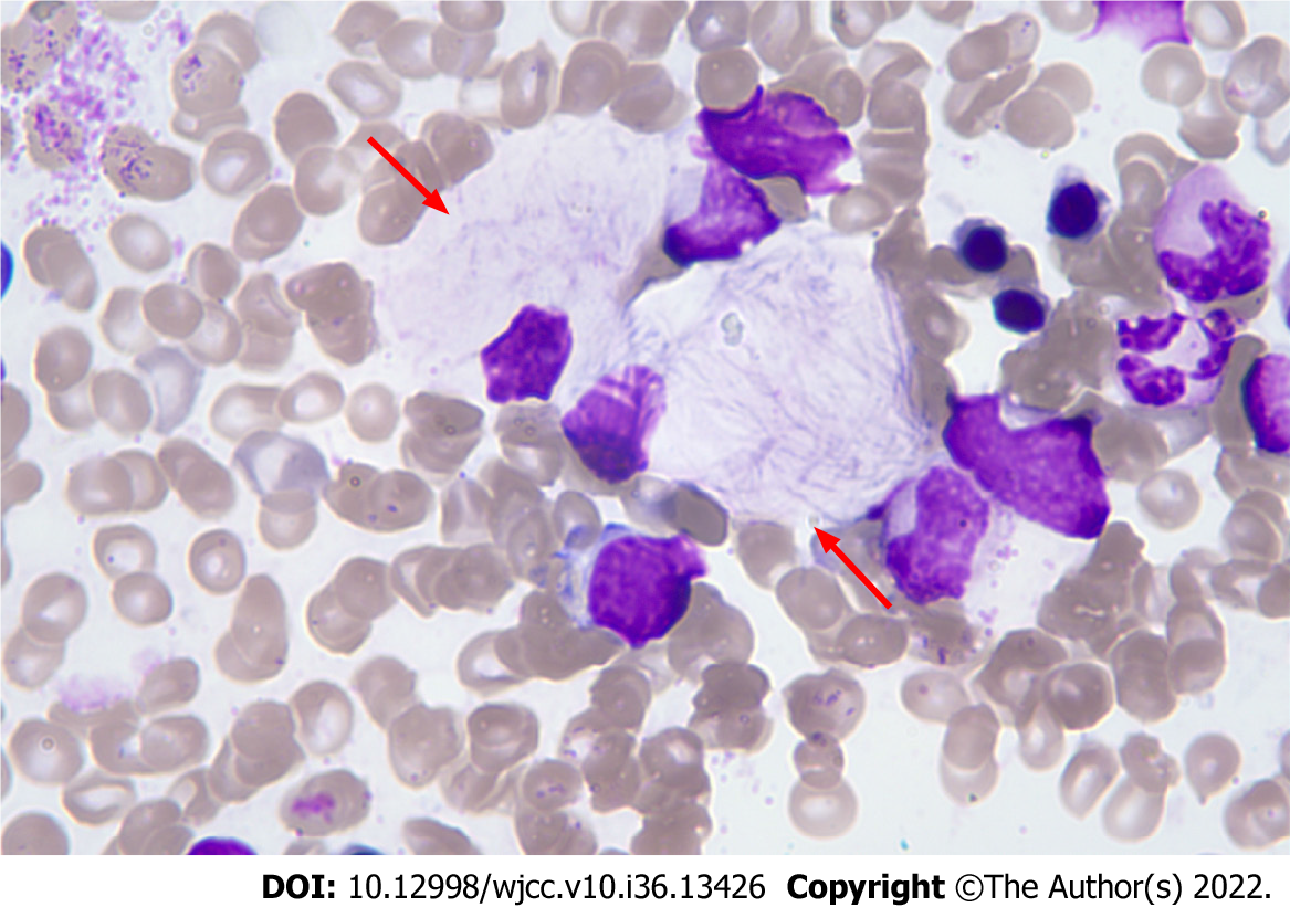
Figure 1 Bone marrow cytomorphology.
Haematoxylin-eosin stain (magnification: 100 ×).
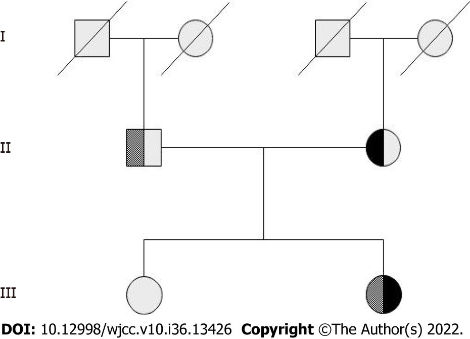
Figure 2 Family pedigree of the proband.
The first-generation members have no information. The second generation is the father of proband is heterozygous for exon 10 c.1448T>C (p.L483P) mutation, and the mother is heterozygous for exon 7 c.928A>G (p.S310G) mutation. The third generation is the proband who was compound heterozygous for p.L483P/p.S310G. Her sister was normal and did not carry either mutation.
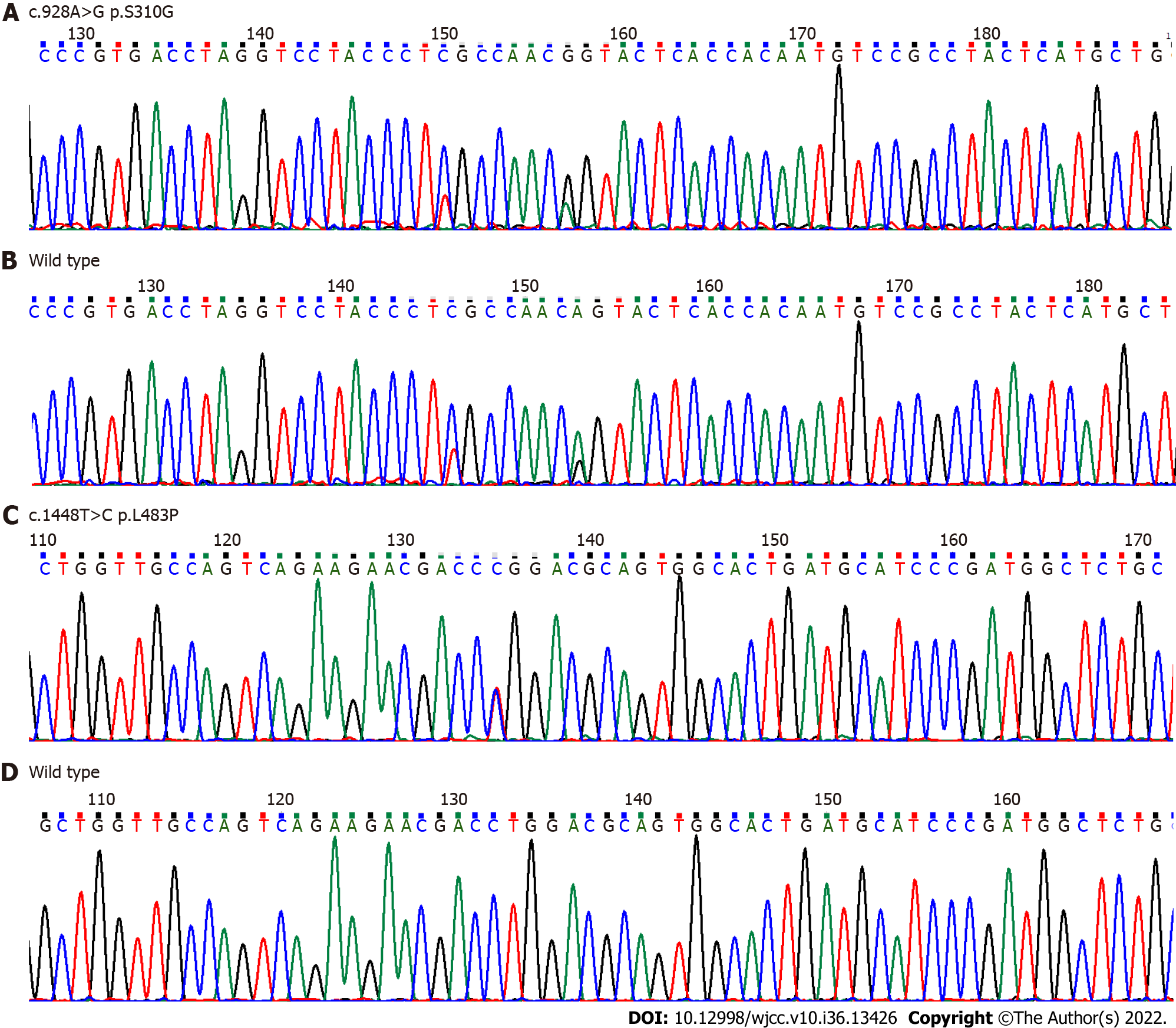
Figure 3 DNA sequencing analysis of glucocerebrosidase gene.
A and B: Exon 7 c.928A>G (p.S310G) novel heterozygous missense mutation compared to the corresponding wild-type sequence; C and D: Exon 10 c.1448T>C (p.L483P) heterozygous mutation compared to the corresponding wild-type sequence.
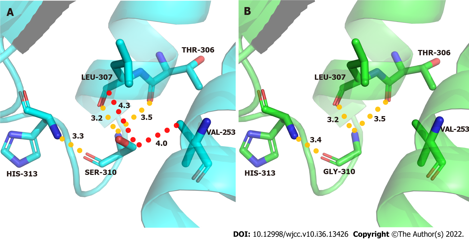
Figure 4 Molecular contacts of residue 310.
A: Wild-type acid β-glucosidase protein; B: Mutant type.
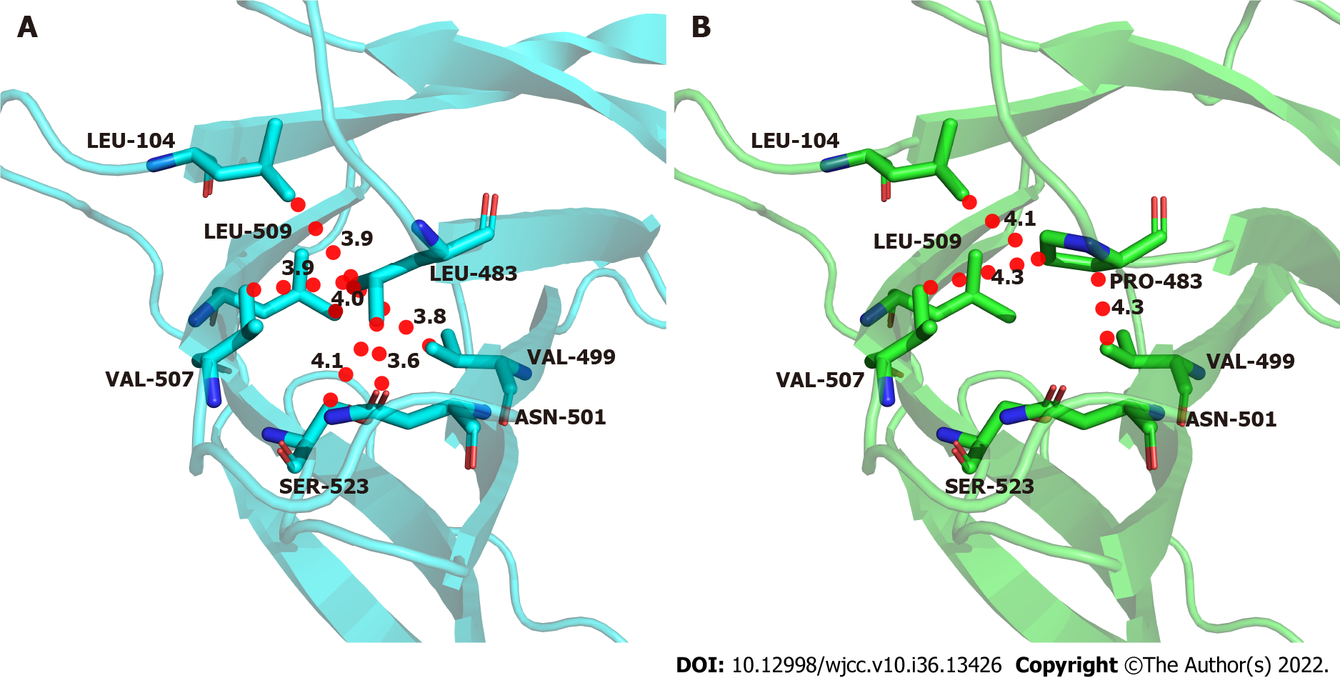
Figure 5 Molecular contacts of residue 483.
A: Wild-type acid β-glucosidase protein; B: Mutant type.
- Citation: Wen XL, Wang YZ, Zhang XL, Tu JQ, Zhang ZJ, Liu XX, Lu HY, Hao GP, Wang XH, Yang LH, Zhang RJ. Compound heterozygous p.L483P and p.S310G mutations in GBA1 cause type 1 adult Gaucher disease: A case report. World J Clin Cases 2022; 10(36): 13426-13434
- URL: https://www.wjgnet.com/2307-8960/full/v10/i36/13426.htm
- DOI: https://dx.doi.org/10.12998/wjcc.v10.i36.13426


