Copyright
©The Author(s) 2022.
World J Clin Cases. Dec 26, 2022; 10(36): 13396-13401
Published online Dec 26, 2022. doi: 10.12998/wjcc.v10.i36.13396
Published online Dec 26, 2022. doi: 10.12998/wjcc.v10.i36.13396
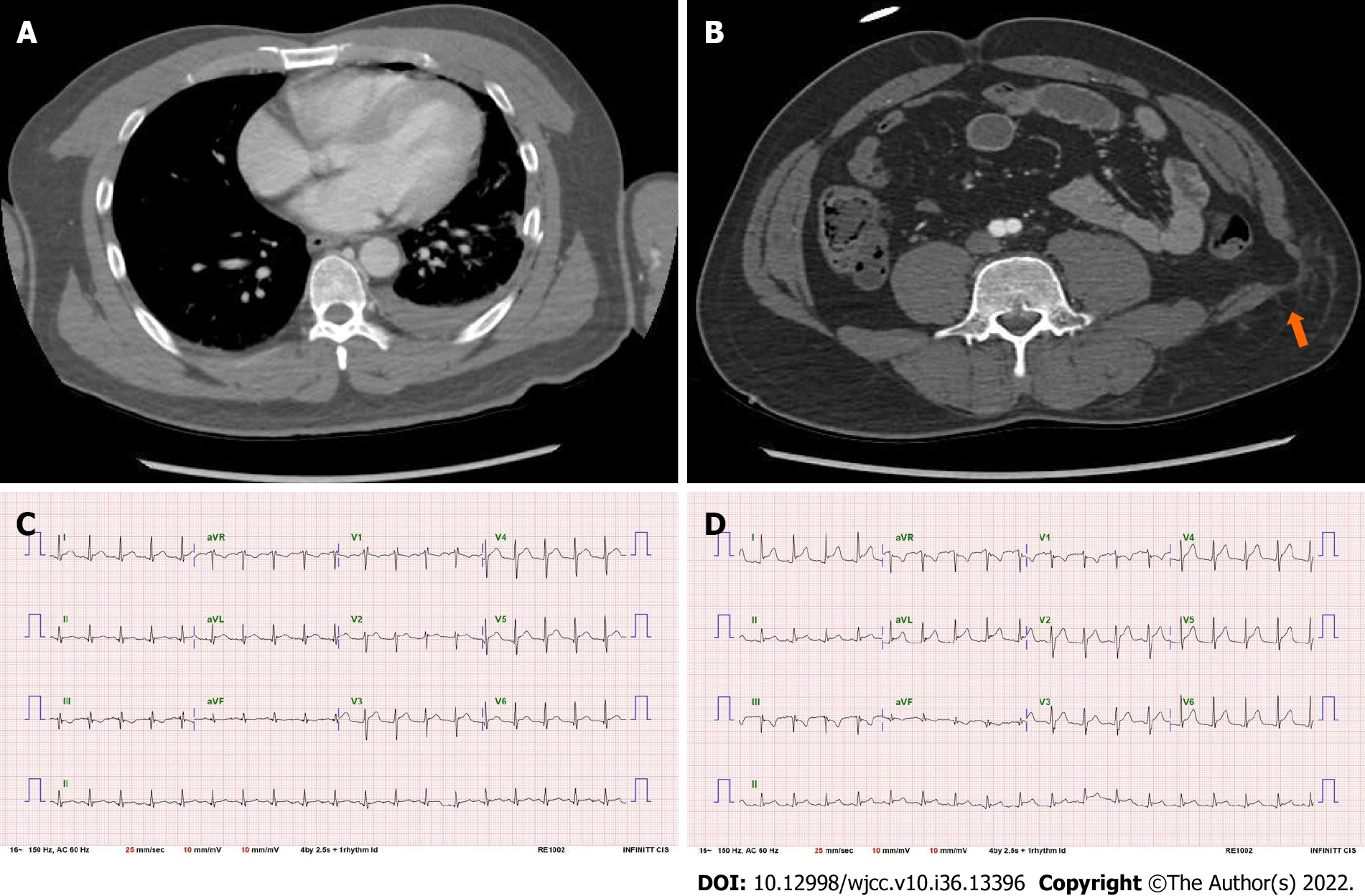
Figure 1 Computed tomography scan.
A: Initial chest contrast computed tomography (CT) showing traumatic hemothorax, with no evidence of heart injury; B: Initial abdominal contrast CT showing visceral herniation through an abdominal wall defect (orange arrow); C: The initial electrocardiogram (ECG) showed widespread abnormal findings, but did not meet the criteria for myocardial infarction (MI); D: ECG performed later when the patient showed fluctuating systolic blood pressure. The ECG showed prominent ST changes with depressions at the lead III, aVR, and V1, which are not the classical reciprocal ECG changes seen in MI.
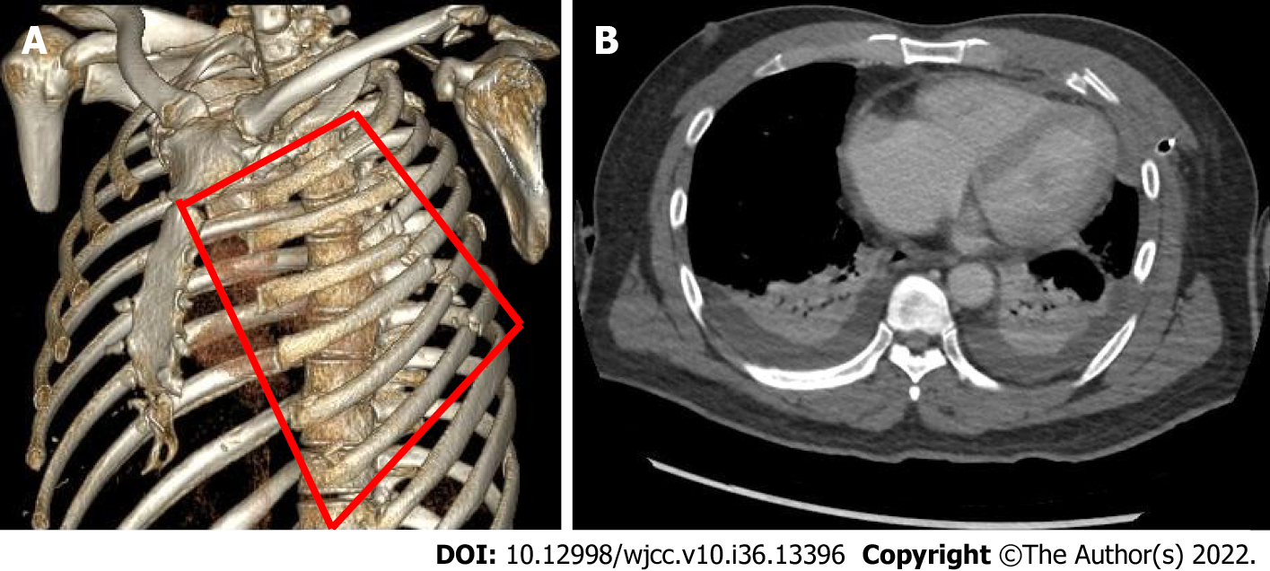
Figure 2 3-dimensional contrast chest computed tomography.
A: A 3-dimensional (3D) contrast chest computed tomography (CT) with rib rendering. The anterolateral flail segment (indicated by the red lines) was located on the left rib cage; B: An axial cut of the 3D chest CT showing bilateral pleural effusion with collapsed lungs, with no evidence of heart injury.
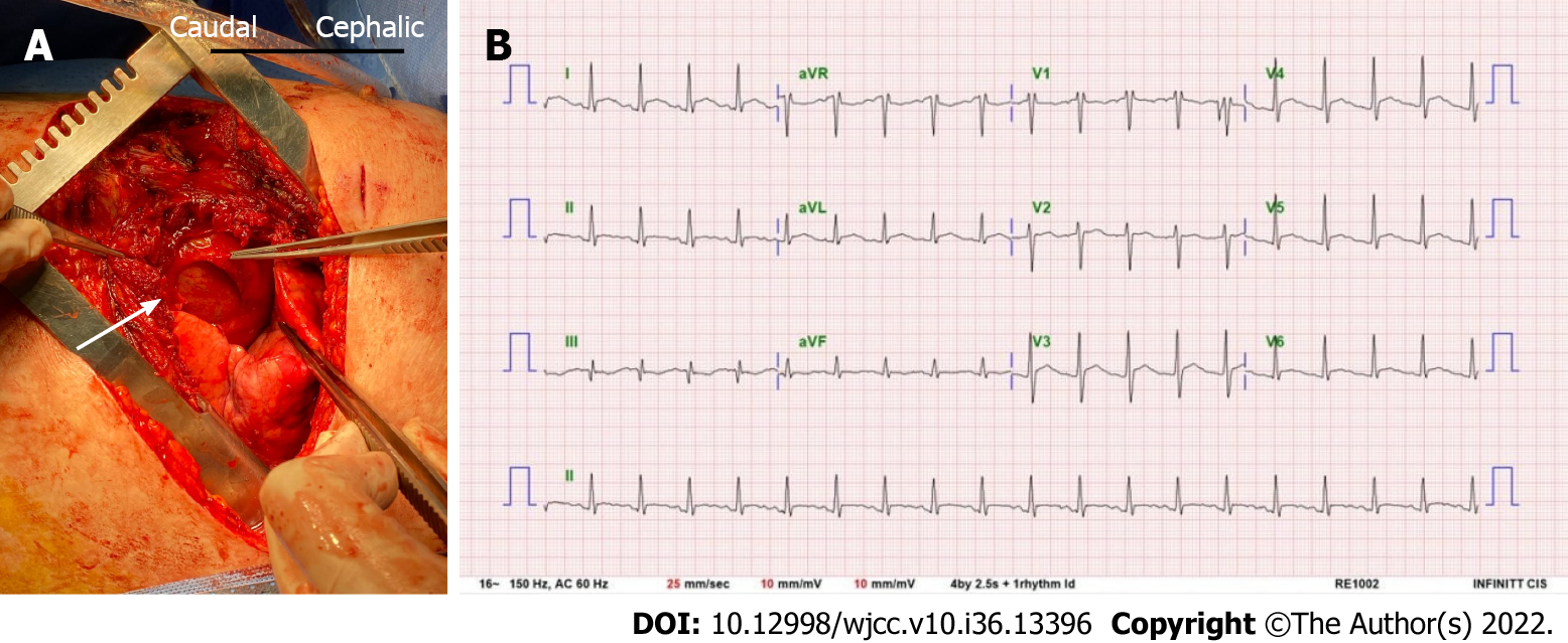
Figure 3 Rib examination.
A: The operation field during surgical stabilization of rib fractures. We routinely performed a thoracic cavity exploration before fixing the rib fractures. The white arrow indicates that the left ventricle was visible through the 4 cm × 4 cm pericardial rupture, which was an incidental finding; B: The postoperative electrocardiogram with the resolution of all ST depressions and lower ST elevations than that present in the preoperative period.
- Citation: Yoon SY, Ye JB, Seok J. Undetected traumatic cardiac herniation like playing hide-and-seek-delayed incidental findings during surgical stabilization of flail chest: A case report. World J Clin Cases 2022; 10(36): 13396-13401
- URL: https://www.wjgnet.com/2307-8960/full/v10/i36/13396.htm
- DOI: https://dx.doi.org/10.12998/wjcc.v10.i36.13396


