Copyright
©The Author(s) 2022.
World J Clin Cases. Dec 26, 2022; 10(36): 13388-13395
Published online Dec 26, 2022. doi: 10.12998/wjcc.v10.i36.13388
Published online Dec 26, 2022. doi: 10.12998/wjcc.v10.i36.13388
Figure 1 Preoperative swelling of both lower extremities and the left thigh.
A: The unequal appearance of the lower limbs; B and C: The inflammatory pseudotumor of the left thigh.
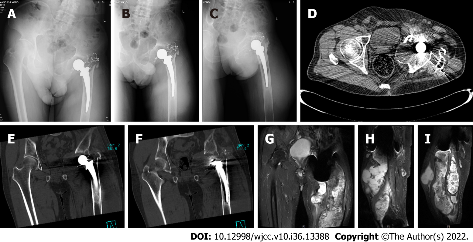
Figure 2 Preoperative imaging examination.
A: X-ray; A-C: Left superior pubic branch - irregular pubic comb bone; D: CT; D-F: Partial bone absorption of the upper part of the left femur; bone destruction of the left acetabulum; swelling and unclear layers of soft tissue shadows in the upper part of the left thigh and around the hip joint; multiple cystic lesions in the upper part of the left thigh, with multiple cystic necrosis areas in the lesions, of which the posterior subcutaneous necrosis area is a long strip from the subcutaneous buttock to the lower back; G: MRI; G-I: The left upper femur and left hip joint have abnormal bone, with multiple necrosis sites around the hip joint and soft tissue of the left thigh as the main cystic disease. Considering the inflammatory disease, the size of the larger cystic lesion is approximately 8.7cm × 9.3 cm × 18 cm.
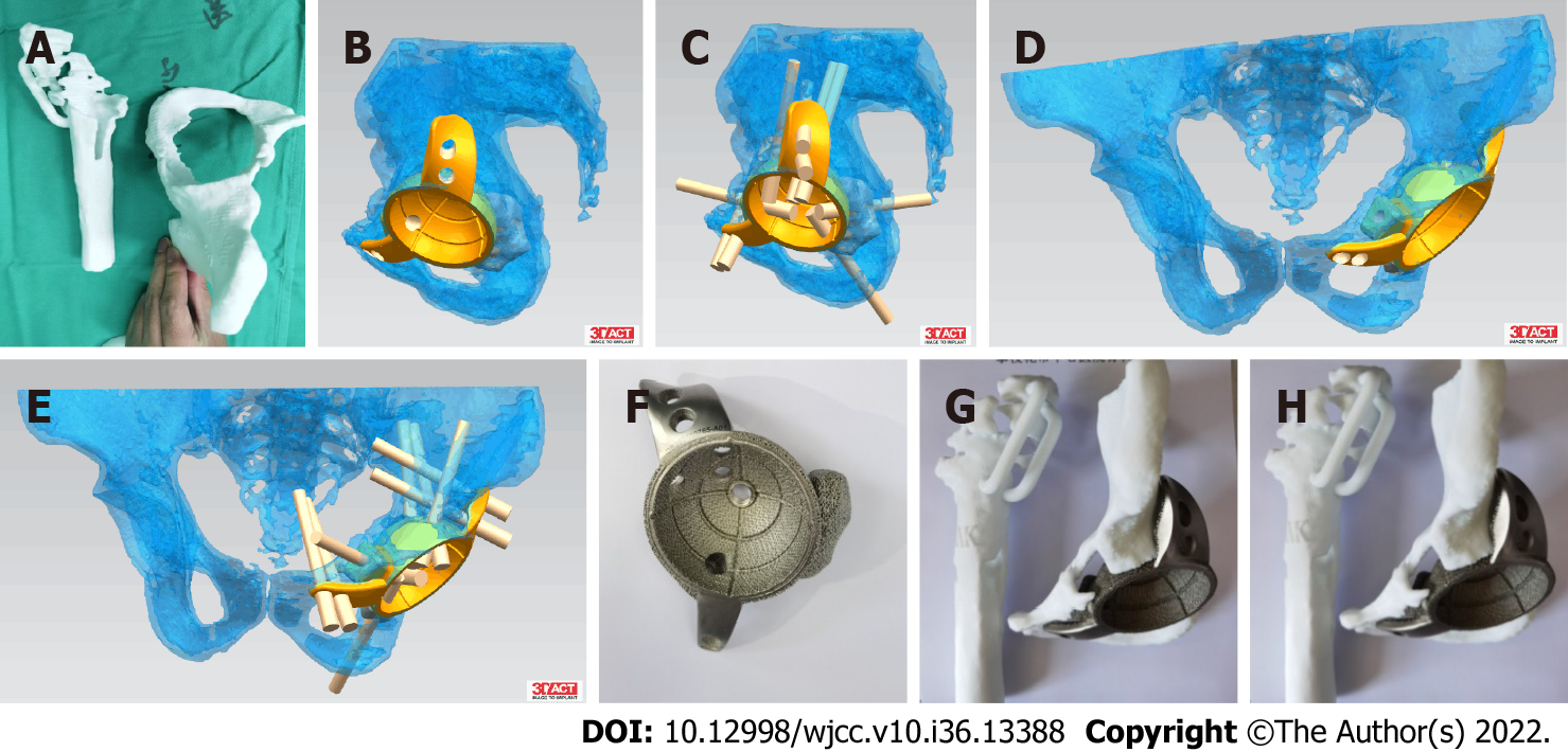
Figure 3 Design process of personalized prosthesis.
A: 1:1 printed model; B-E: Preoperative planning; F: Cup making; G-H: Preoperative surgery simulation.
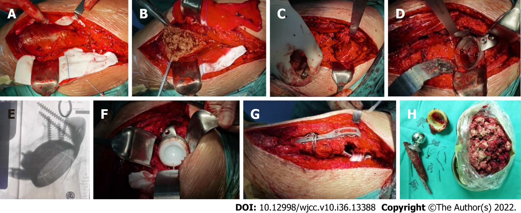
Figure 4 Surgical steps.
A: The inflammatory pseudotumor was freed; B: The pseudotumor was excised; C. Acetabular defect; D. Implanted acetabular cup; E. Intraoperative fluoroscopy; F. Uchimura implant; G. Steel plate-fixed rotor; H. Removal of prosthesis and inflammatory pseudotumor.
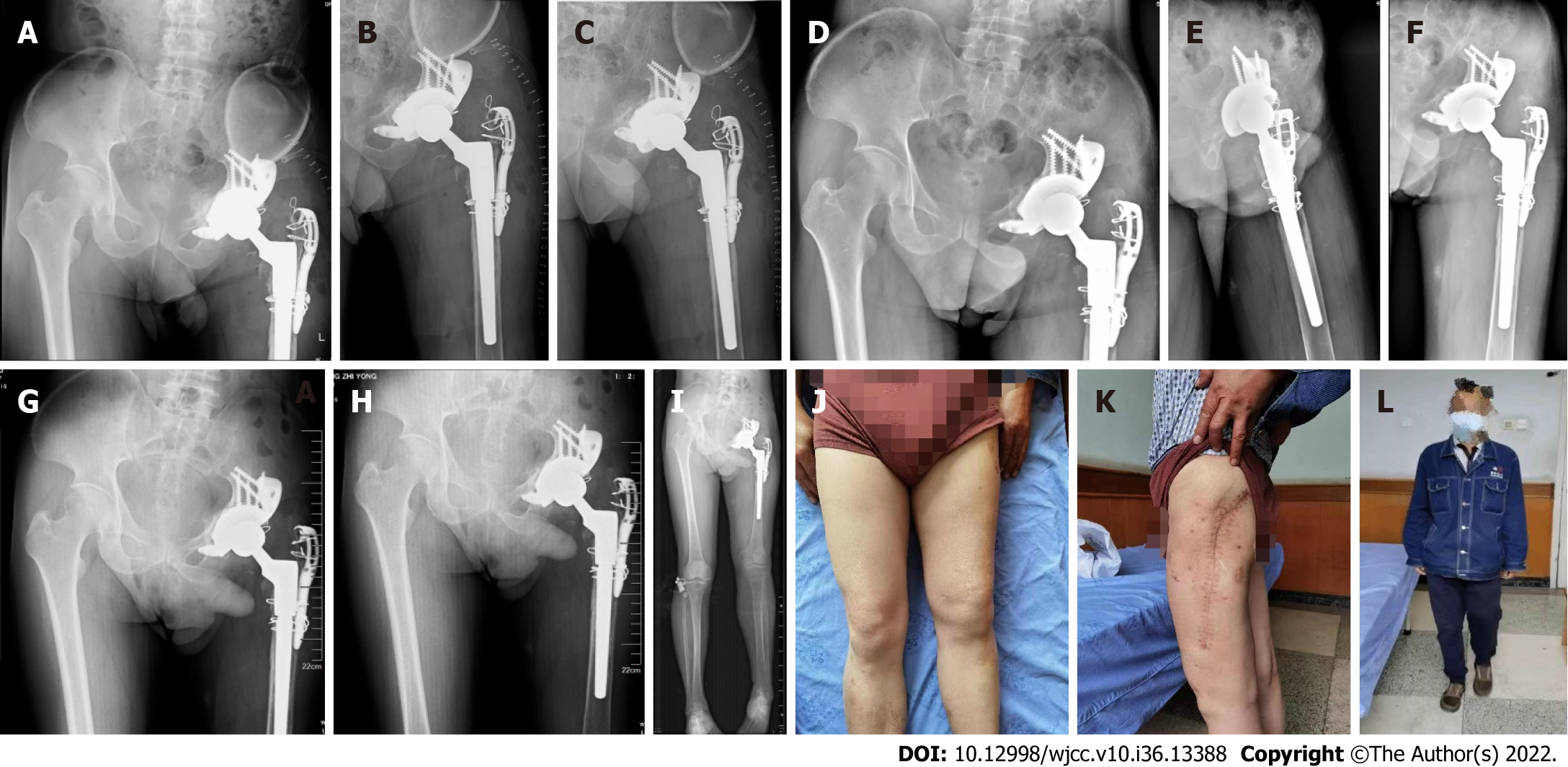
Figure 5 Postoperative follow-up.
A: X-ray in two days after surgery; B and C: Different view; D-F: Ninety days after surgery; G-L: Six months after surgery (a 30-cm surgical scar on the outside of the left thigh, without redness, swelling or exudation).
- Citation: Wang HP, Wang MY, Lan YP, Tang ZD, Tao QF, Chen CY. Application of 3D-printed prosthesis in revision surgery with large inflammatory pseudotumour and extensive bone defect: A case report. World J Clin Cases 2022; 10(36): 13388-13395
- URL: https://www.wjgnet.com/2307-8960/full/v10/i36/13388.htm
- DOI: https://dx.doi.org/10.12998/wjcc.v10.i36.13388


