Copyright
©The Author(s) 2022.
World J Clin Cases. Dec 26, 2022; 10(36): 13356-13363
Published online Dec 26, 2022. doi: 10.12998/wjcc.v10.i36.13356
Published online Dec 26, 2022. doi: 10.12998/wjcc.v10.i36.13356
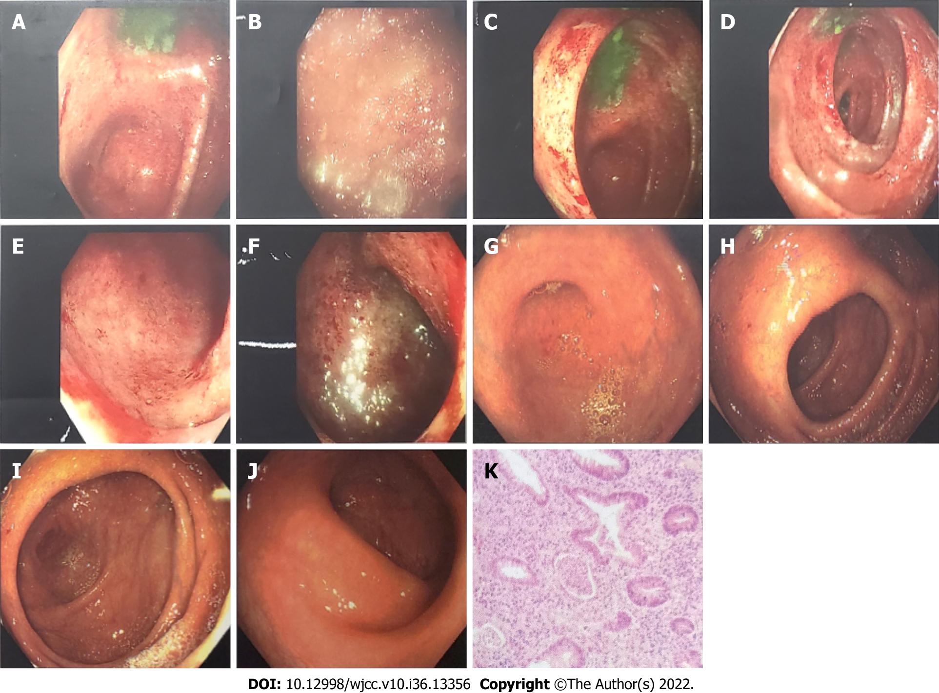
Figure 1 Comparison of colonoscopy results before and after treatment.
A-F: Before treatment (January 19, 2018): The mucosa of the whole colon and rectum was congested and edematous, showing patchy hemorrhagic foci, with purulent moss on the surface and partial erosion, of which the right colon and rectum were the most serious; G-J: After treatment (September 9, 2018): There was no hyperemia or swelling around the appendix, clear hyperemia or edema of the mucosa of the blind base and ascending colon, white mucus on the surface, erosion or bleeding, and no erosion or bleeding of the mucosa of the liver flexure, transverse colon, splenic flexure, descending colon and sigmoid colon, ulcers or new organisms were observed. The rectal mucosa was free of erosion, bleeding and new organisms; K: Pathological diagnosis: (Large intestine) mucosa acute and chronic inflammation with crypt abscess formation, consistent with ulcerative colorectal inflammation.
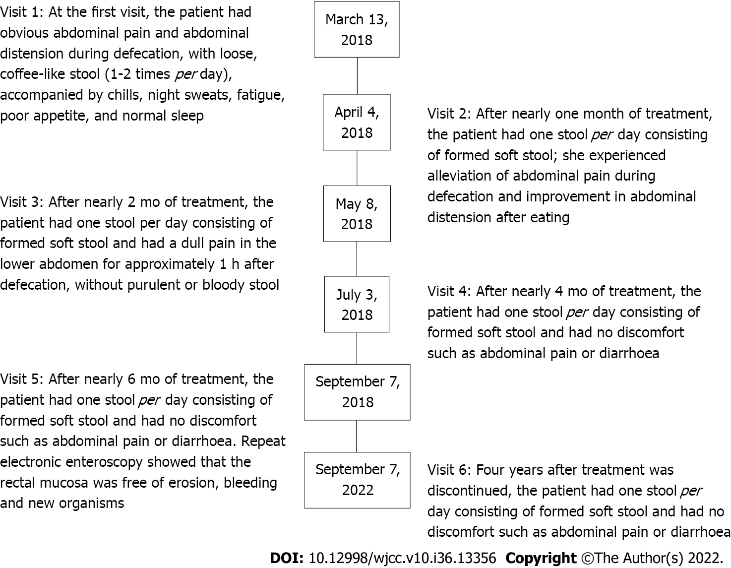
Figure 2 Description of the medical records.
- Citation: Wu B. Acute moderate to severe ulcerative colitis treated by traditional Chinese medicine: A case report. World J Clin Cases 2022; 10(36): 13356-13363
- URL: https://www.wjgnet.com/2307-8960/full/v10/i36/13356.htm
- DOI: https://dx.doi.org/10.12998/wjcc.v10.i36.13356


