Copyright
©The Author(s) 2022.
World J Clin Cases. Dec 26, 2022; 10(36): 13179-13188
Published online Dec 26, 2022. doi: 10.12998/wjcc.v10.i36.13179
Published online Dec 26, 2022. doi: 10.12998/wjcc.v10.i36.13179
Figure 1 Comparison of the cortical bone trajectory screw with the traditional pedicle screw trajectory.
A: Axial view; B: Lateral view; C: Anteroposterior view.
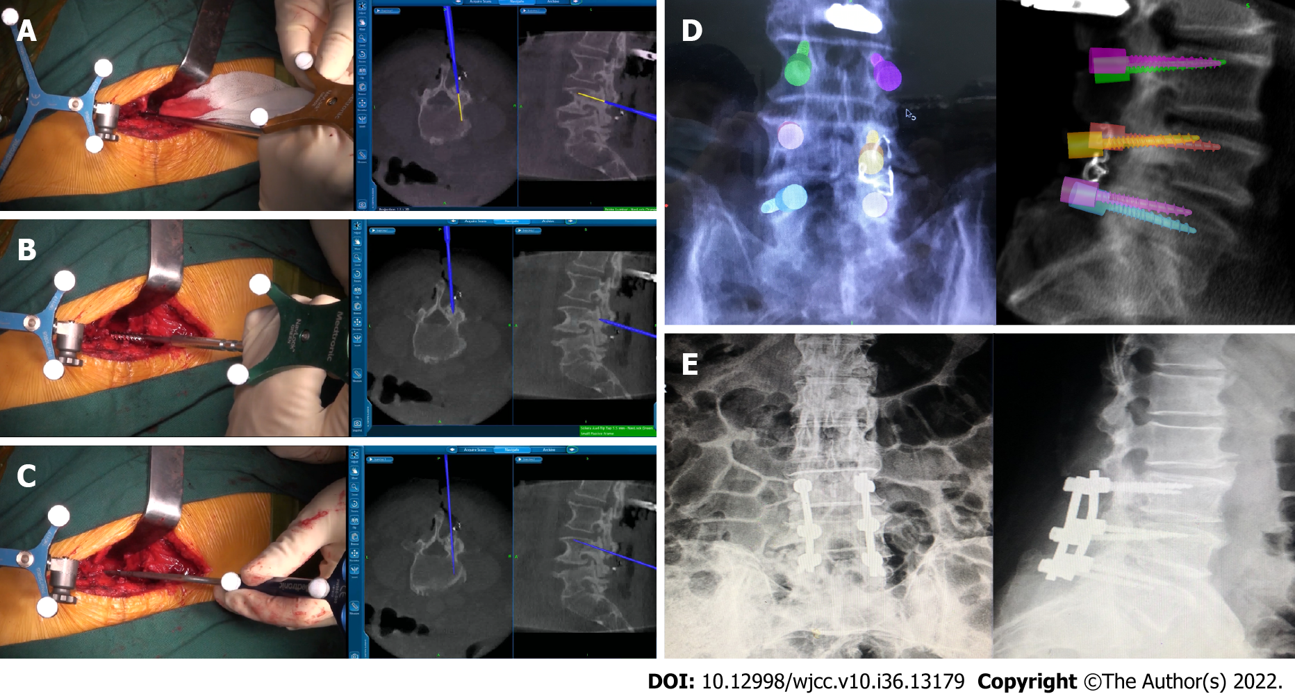
Figure 2 Implantation of the cortical bone trajectory screw assisted by the navigation system.
A: Feeling the entry point of the cortical bone trajectory (CBT) screw in L4 with the assistance of the navigation system; B: Awl of the CBT screw in L4 with the assistance of the navigation system; C: Tapping of the CBT screw in L4 with the assistance of the navigation system; D: Fluoroscopy showed the placement of CBT screws during the surgery; E: X-ray showed the implantation of CBT screws after surgery.
- Citation: Guo S, Zhu K, Yan MJ, Li XH, Tan J. Cortical bone trajectory screws in the treatment of lumbar degenerative disc disease in patients with osteoporosis. World J Clin Cases 2022; 10(36): 13179-13188
- URL: https://www.wjgnet.com/2307-8960/full/v10/i36/13179.htm
- DOI: https://dx.doi.org/10.12998/wjcc.v10.i36.13179


