Copyright
©The Author(s) 2022.
World J Clin Cases. Dec 6, 2022; 10(34): 12775-12780
Published online Dec 6, 2022. doi: 10.12998/wjcc.v10.i34.12775
Published online Dec 6, 2022. doi: 10.12998/wjcc.v10.i34.12775
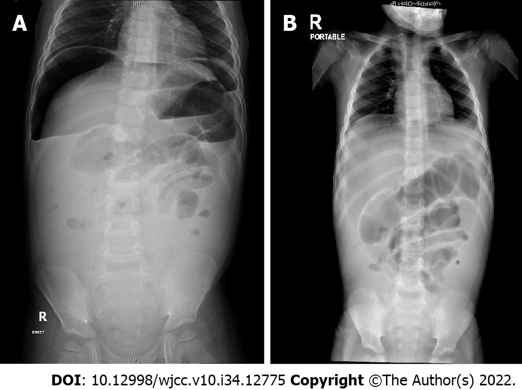
Figure 1 Abdominal X-ray demonstrated a significant amount of pneumoperitoneum.
A: Supine view; B: Erect view.
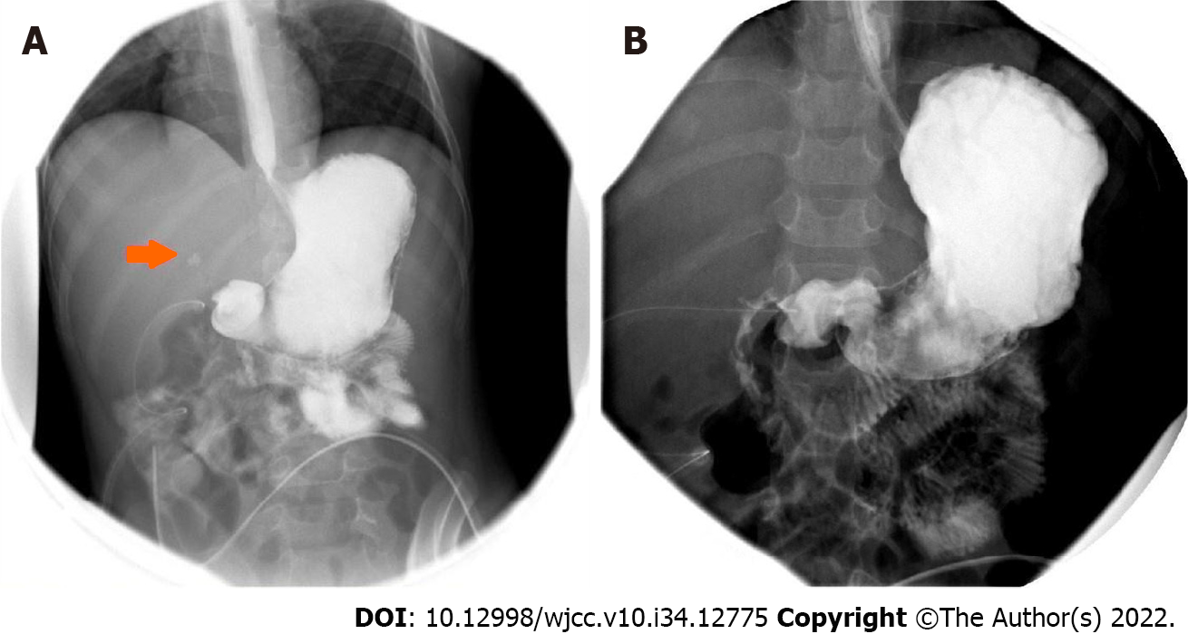
Figure 2 Upper gastrointestinal study.
A: Upper gastrointestinal study with water-soluble contrast showed minimal leakage in the first part of the duodenum. Two abdominal drains were also visualized; B: Repeated upper gastrointestinal study 2 wk after surgery showed no evidence of contrast leakage.
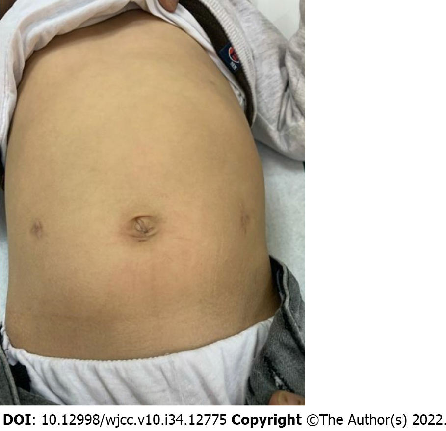
Figure 3
The patient had satisfactory cosmetic results at the follow-up.
- Citation: Alshehri A, Alsinan TA. Perforated duodenal ulcer secondary to deferasirox use in a child successfully managed with laparoscopic drainage: A case report . World J Clin Cases 2022; 10(34): 12775-12780
- URL: https://www.wjgnet.com/2307-8960/full/v10/i34/12775.htm
- DOI: https://dx.doi.org/10.12998/wjcc.v10.i34.12775


