Copyright
©The Author(s) 2022.
World J Clin Cases. Dec 6, 2022; 10(34): 12717-12725
Published online Dec 6, 2022. doi: 10.12998/wjcc.v10.i34.12717
Published online Dec 6, 2022. doi: 10.12998/wjcc.v10.i34.12717
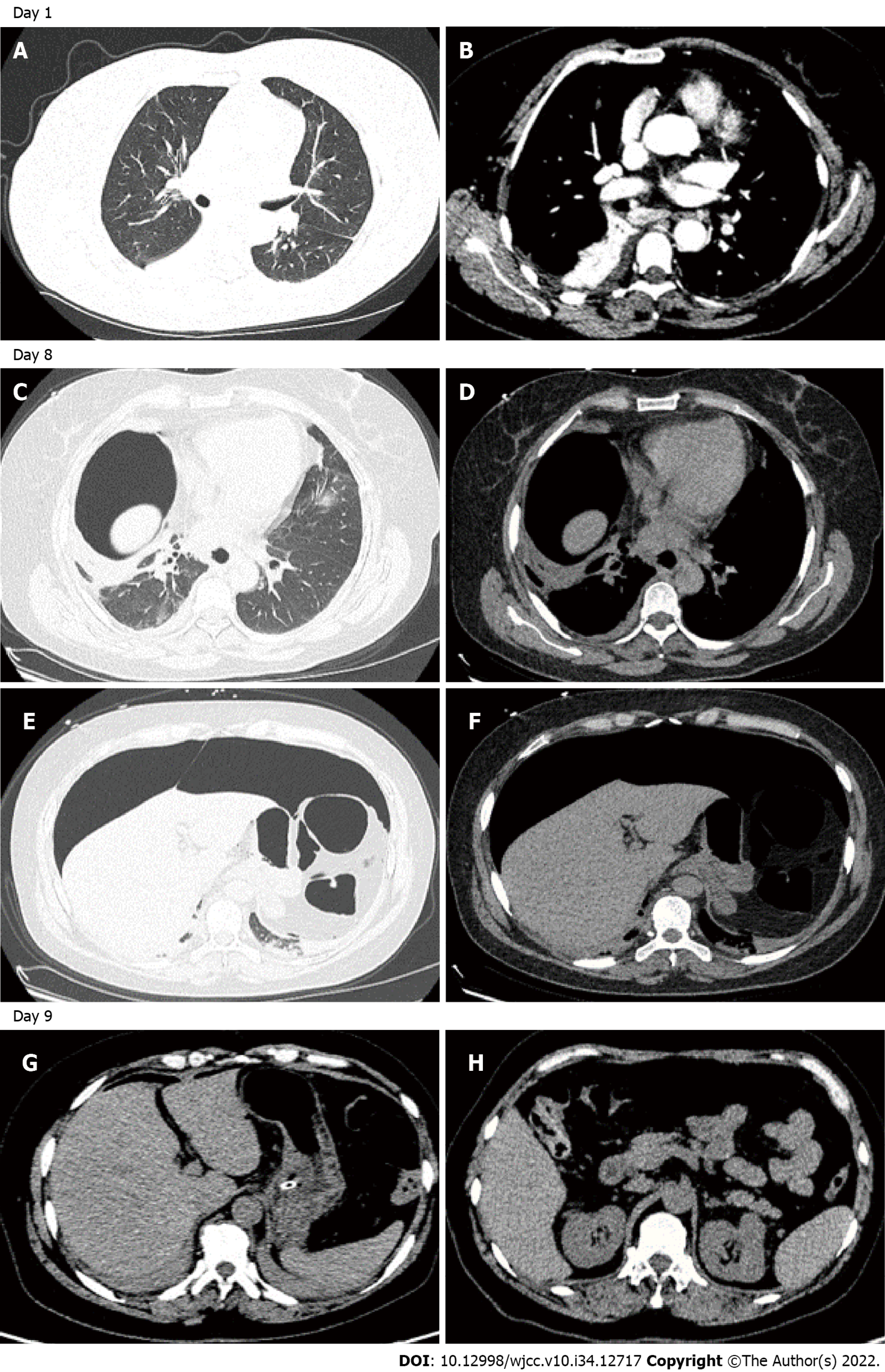
Figure 1 Chest and abdomenal computed tomography scan of the patient on day 1, day 8, and day 9.
A and B: Thoracic enhanced computed tomography (CT)-scan on admission showed infiltration and fibrotic foci were scattered in the bilateral pulmonary lobe, with localized consolidation of the right lower lobe, and bilateral pleural effusions; C-F: Thoracic and abdominal CT-scan on day 8 taken after needle decompression demonstrating the presence of a large anterior pneumoperitoneum without fluid collections and pneumothorax or pneumomediastinium; G and H: Abdominal enhanced CT-scan on day 9 showed gas accumulation in the right abdominal wall, free gas in the abdominal cavity is less than in the previous CT exam.
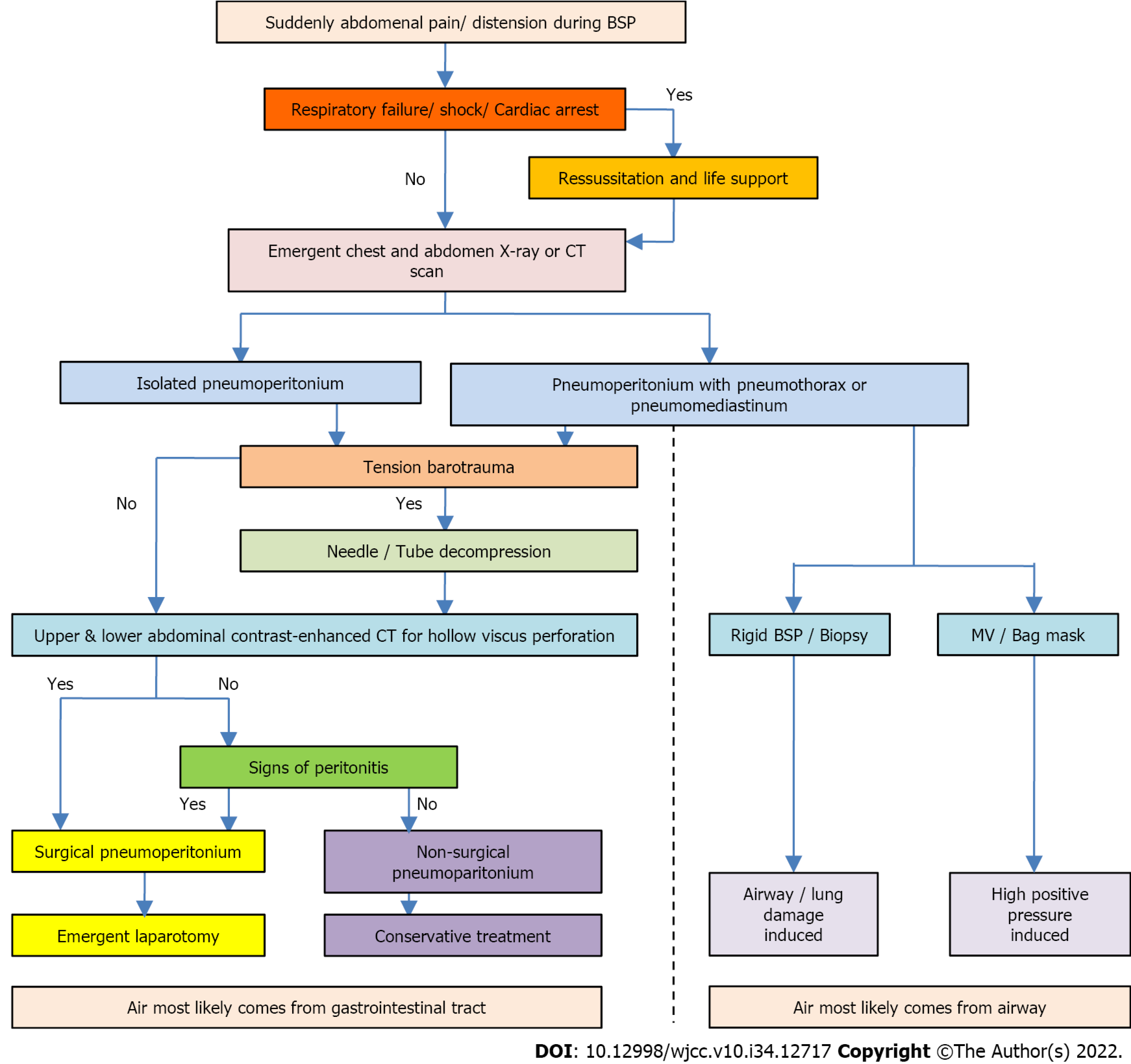
Figure 2 Protocol for the management of pneumoperitonium during bronchoscopy.
BSP: Bronchoscopy; MV: Mechanical ventilation; CT: Computed tomography.
- Citation: Baima YJ, Shi DD, Shi XY, Yang L, Zhang YT, Xiao BS, Wang HY, He HY. How to manage isolated tension non-surgical pneumoperitonium during bronchoscopy? A case report . World J Clin Cases 2022; 10(34): 12717-12725
- URL: https://www.wjgnet.com/2307-8960/full/v10/i34/12717.htm
- DOI: https://dx.doi.org/10.12998/wjcc.v10.i34.12717


