Copyright
©The Author(s) 2022.
World J Clin Cases. Dec 6, 2022; 10(34): 12696-12702
Published online Dec 6, 2022. doi: 10.12998/wjcc.v10.i34.12696
Published online Dec 6, 2022. doi: 10.12998/wjcc.v10.i34.12696
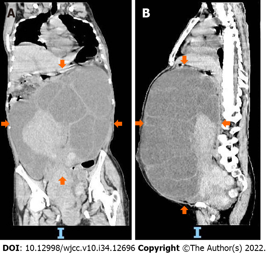
Figure 1 Preoperative computed tomography images of the patient.
A and B: Computed tomography shows a large cystic solid mass in the pelvic and abdominal cavity (orange arrows).
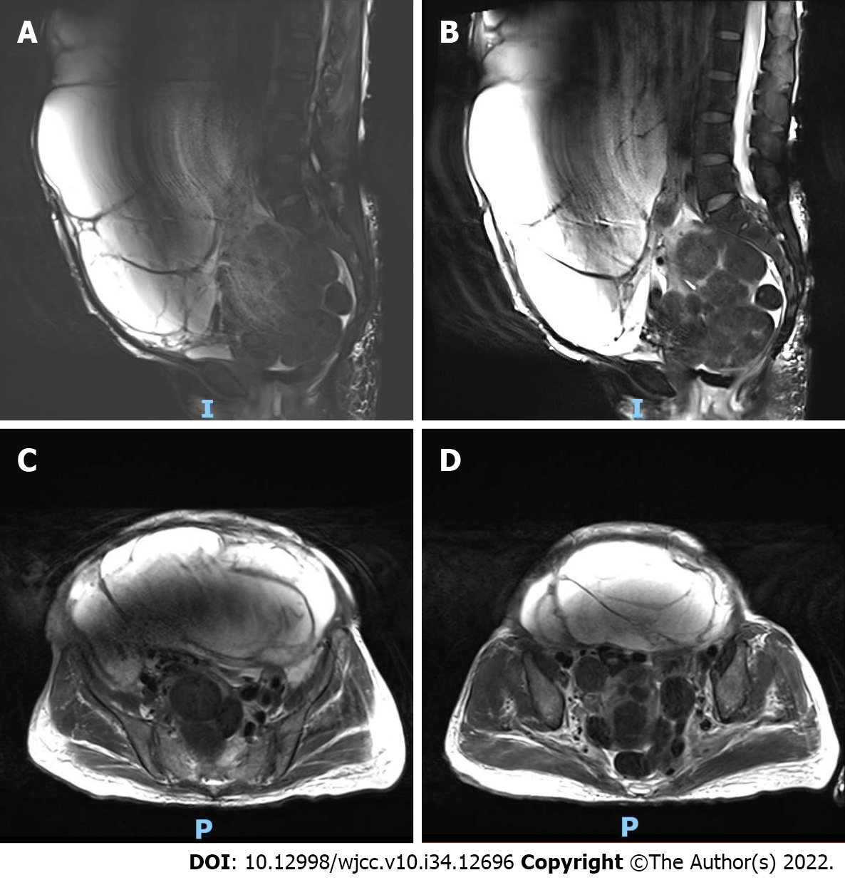
Figure 2 Preoperative magnetic resonance imaging of the mass.
A and B: Magnetic resonance imaging of a large cystic solid mass observed in the abdomen and pelvis, approximately 32.0 cm × 21.1 cm × 37.4 cm in size. Slightly longer T1 and slightly shorter T2 signal changes in the solid component, and multiple cystic changes, long T1 and long T2 signal shadow in the cystic component; C and D: Magnetic resonance imaging of the smooth cyst wall of the lesion, and the clear boundary between the lesion and the surrounding tissue structure. The compressed and displaced abdominal and pelvic organs are shown.
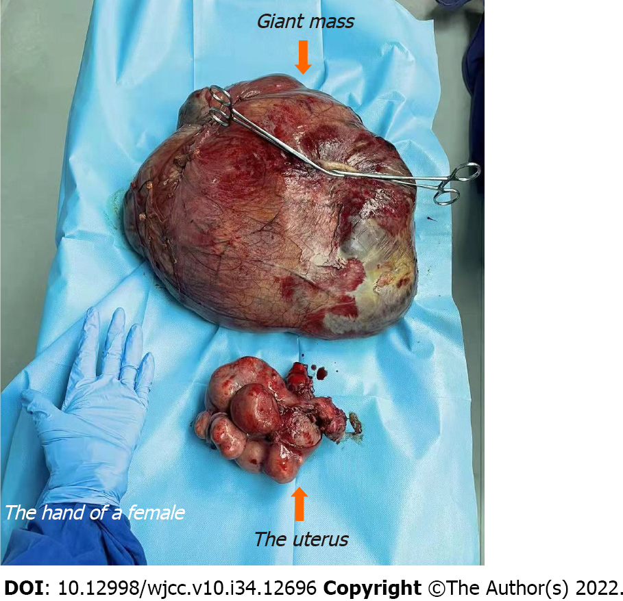
Figure 3 Postoperative mass and uterus (orange arrows).
More than 10 masses were seen on the uterus surface and a large mass was removed from the right broad ligament.
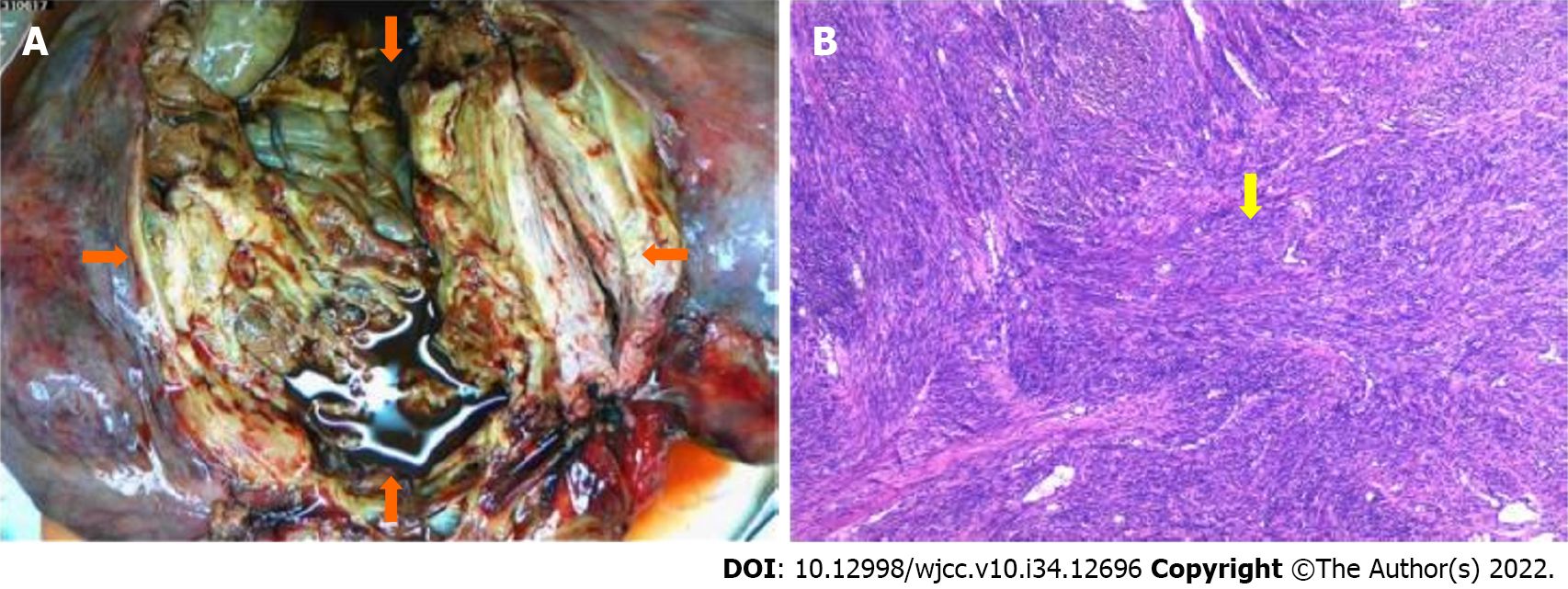
Figure 4 The tumor after surgical dissection (orange arrows).
A and B: Uterine masses were composed of smooth muscle cells with proliferation and braided arrangement. The cell morphology was slightly changed and no abnormalities were observed (yellow arrow).
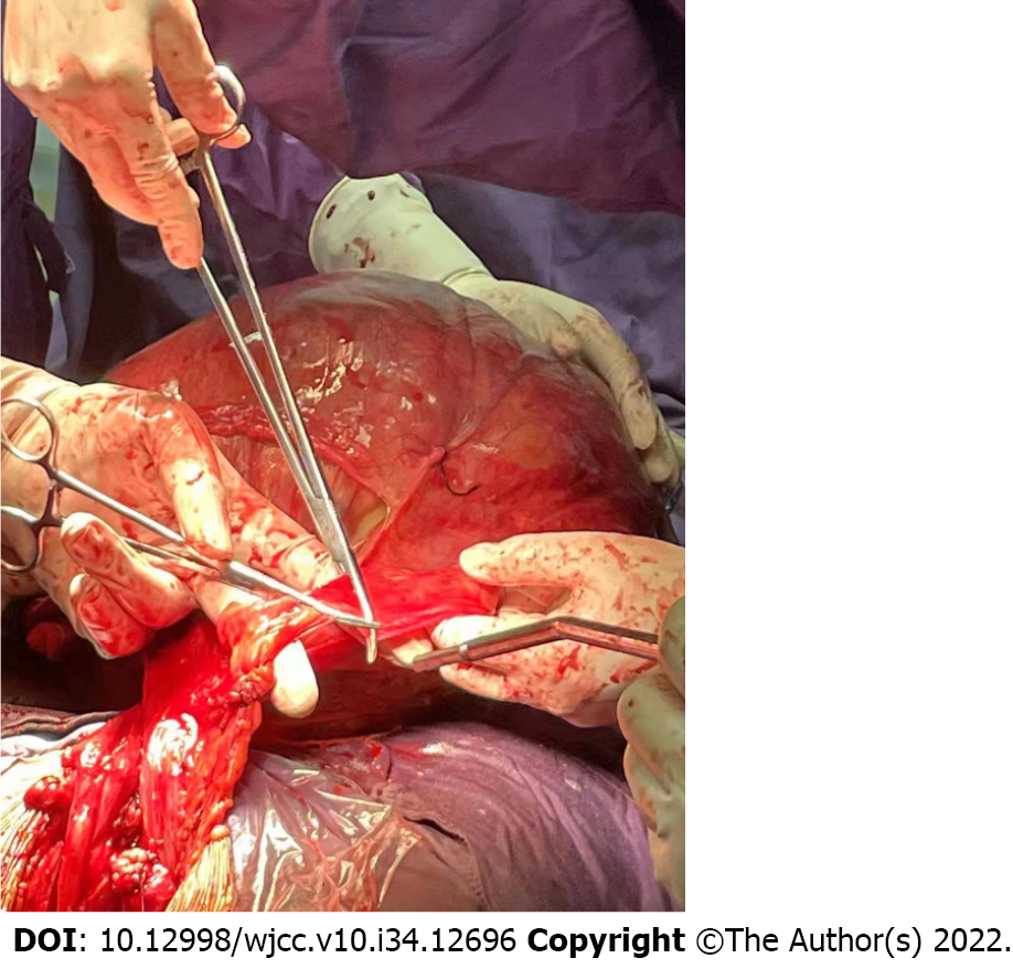
Figure 5 Intraoperative picture of the operation.
The large leiomyoma in the right broad ligament is shown.
- Citation: Yan J, Li Y, Long XY, Li DC, Li SJ. Giant cellular leiomyoma in the broad ligament of the uterus: A case report. World J Clin Cases 2022; 10(34): 12696-12702
- URL: https://www.wjgnet.com/2307-8960/full/v10/i34/12696.htm
- DOI: https://dx.doi.org/10.12998/wjcc.v10.i34.12696


