Copyright
©The Author(s) 2022.
World J Clin Cases. Dec 6, 2022; 10(34): 12690-12695
Published online Dec 6, 2022. doi: 10.12998/wjcc.v10.i34.12690
Published online Dec 6, 2022. doi: 10.12998/wjcc.v10.i34.12690
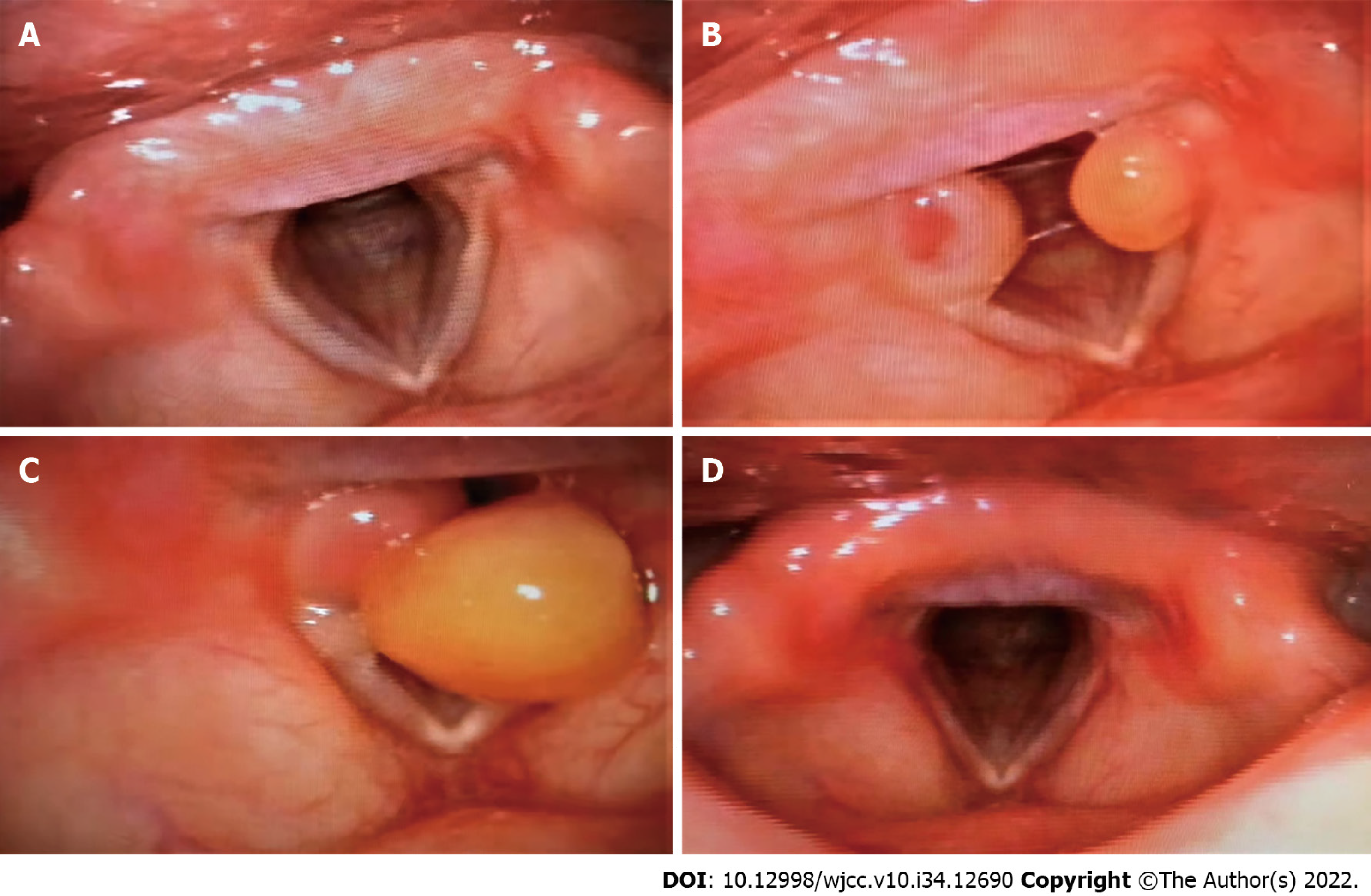
Figure 1 Fibrolaryngoscopy changes of the glottis before and after thoracic surgery.
A: Fibrolaryngoscopy 6 mo before thoracic surgery showed no neoplasm at the glottis; B: Fibrolaryngoscopy 51 d after thoracic surgery revealed granulation tissue in the bilateral arytenoid region of vocal cords; C: Fibrolaryngoscopy 73 d after thoracic surgery revealed granulation tissue in the bilateral arytenoid region of vocal cords; D: Fibrolaryngoscopy 105 d after thoracic surgery revealed no granulation tissue in the bilateral arytenoid region of vocal cords.
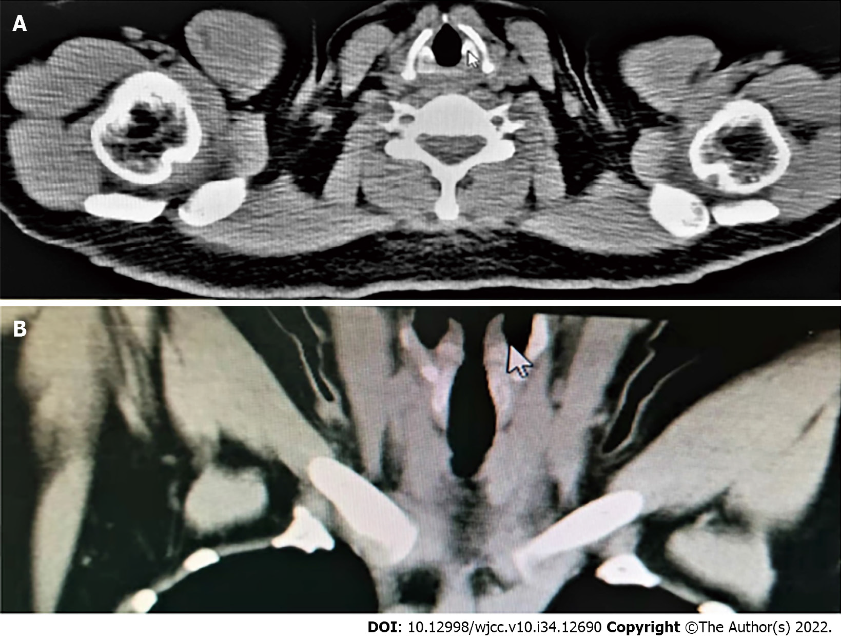
Figure 2 No neoplasm at the glottis was observed on chest computed tomography during thoracic surgery.
A: Cross-section or longitudinal chest computed tomography; B: Longitudinal chest computed tomography.
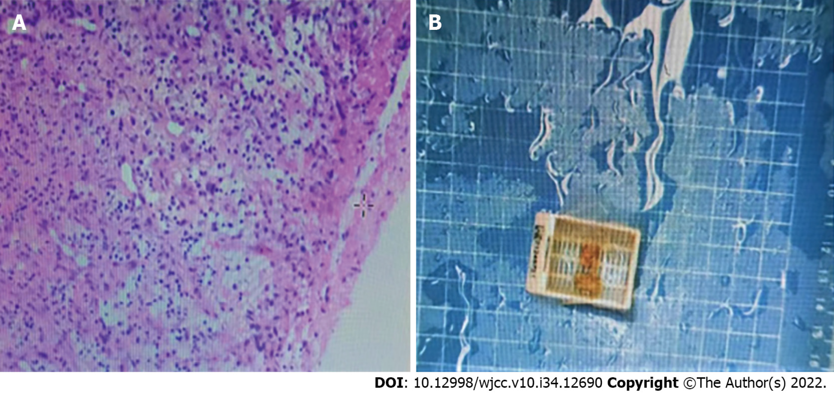
Figure 3 Pathological report after vocal cord polypectomy showed bilateral vocal cord polyps with a large amount of granulation tissue in the stroma.
A: Pathological section diagram; B: Pathological examination (hematoxylin-eosin staining).
- Citation: Xiong XJ, Wang L, Li T. Formation of granulation tissue on bilateral vocal cords after double-lumen endotracheal intubation: A case report. World J Clin Cases 2022; 10(34): 12690-12695
- URL: https://www.wjgnet.com/2307-8960/full/v10/i34/12690.htm
- DOI: https://dx.doi.org/10.12998/wjcc.v10.i34.12690


