Copyright
©The Author(s) 2022.
World J Clin Cases. Dec 6, 2022; 10(34): 12678-12683
Published online Dec 6, 2022. doi: 10.12998/wjcc.v10.i34.12678
Published online Dec 6, 2022. doi: 10.12998/wjcc.v10.i34.12678
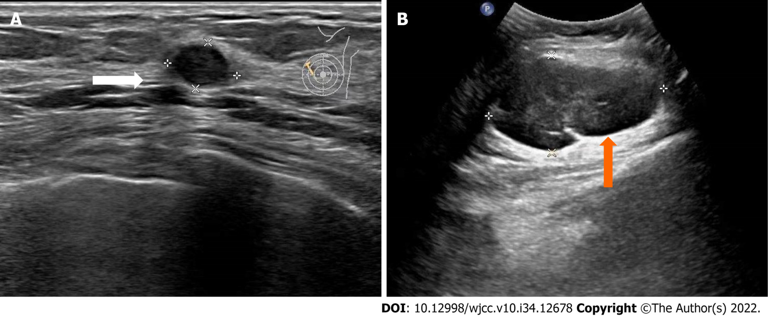
Figure 1 Color ultrasonography of the breast and axilla.
A: Low-echo nodules in the left breast (white arrow); B: Mass in the left axilla (orange arrow).
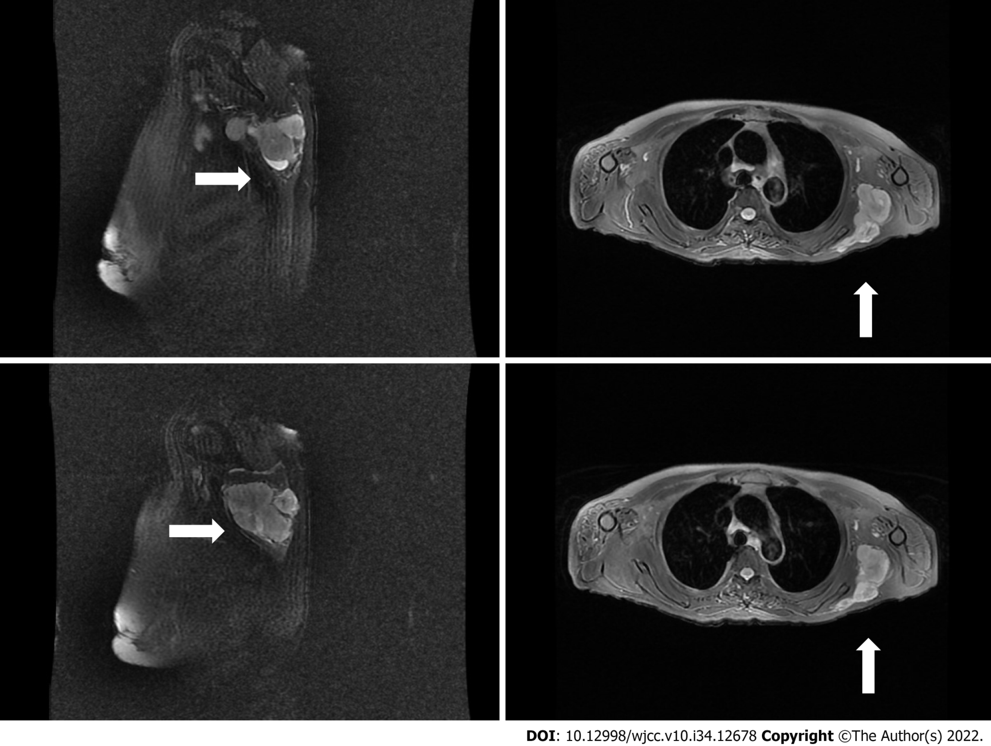
Figure 2 Magnetic resonance imaging of the shoulder joint.
Mass in the left dorsi muscle (white arrow) was observed.
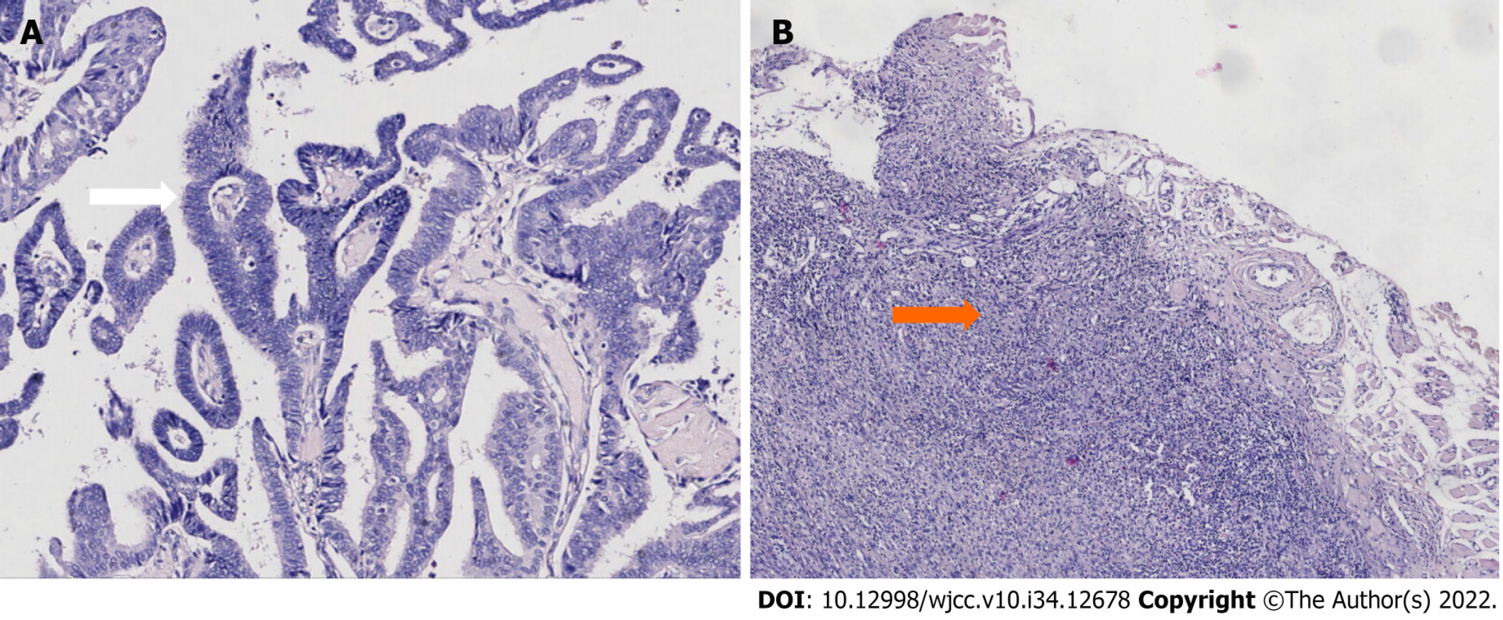
Figure 3 Histopathological examination.
Left breast cancer (white arrow) and left axillary sarcoma (orange arrow). A: Hematoxylin-eosin staining (× 100); B: Hematoxylin-eosin staining (× 40).
- Citation: Gao N, Yang AQ, Xu HR, Li L. Malignant fibrous histiocytoma of the axilla with breast cancer: A case report. World J Clin Cases 2022; 10(34): 12678-12683
- URL: https://www.wjgnet.com/2307-8960/full/v10/i34/12678.htm
- DOI: https://dx.doi.org/10.12998/wjcc.v10.i34.12678


