Copyright
©The Author(s) 2022.
World J Clin Cases. Dec 6, 2022; 10(34): 12671-12677
Published online Dec 6, 2022. doi: 10.12998/wjcc.v10.i34.12671
Published online Dec 6, 2022. doi: 10.12998/wjcc.v10.i34.12671
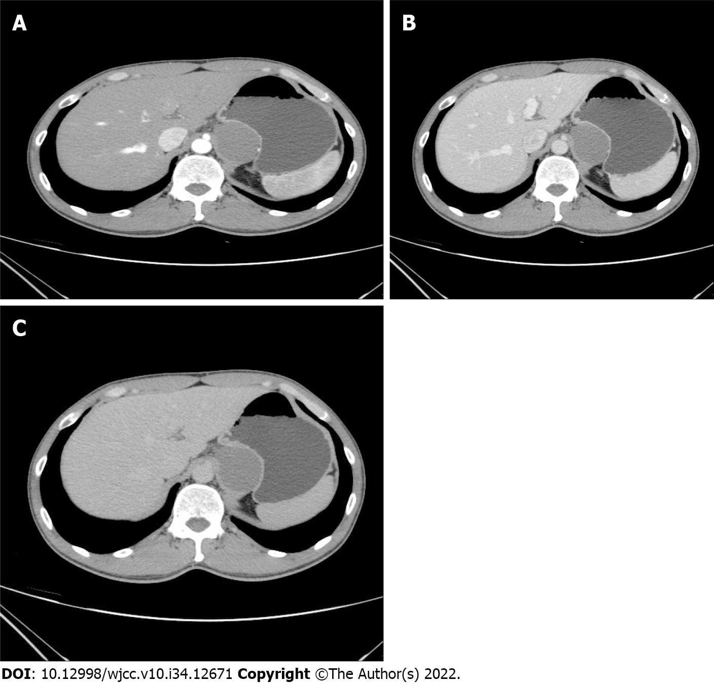
Figure 1 Arterial, venous, and delayed phases.
A: CT value was 44 HU; B: CT value was 39 HU; C: CT value was 35 HU. The lesion had a well-defined margin and was adhered to the posterior wall of the gastric cardia and the inner wall of the gastric fundus, adjacent to the gastric mucosal lining with visible surrounding fat space. CT: Computed tomography.
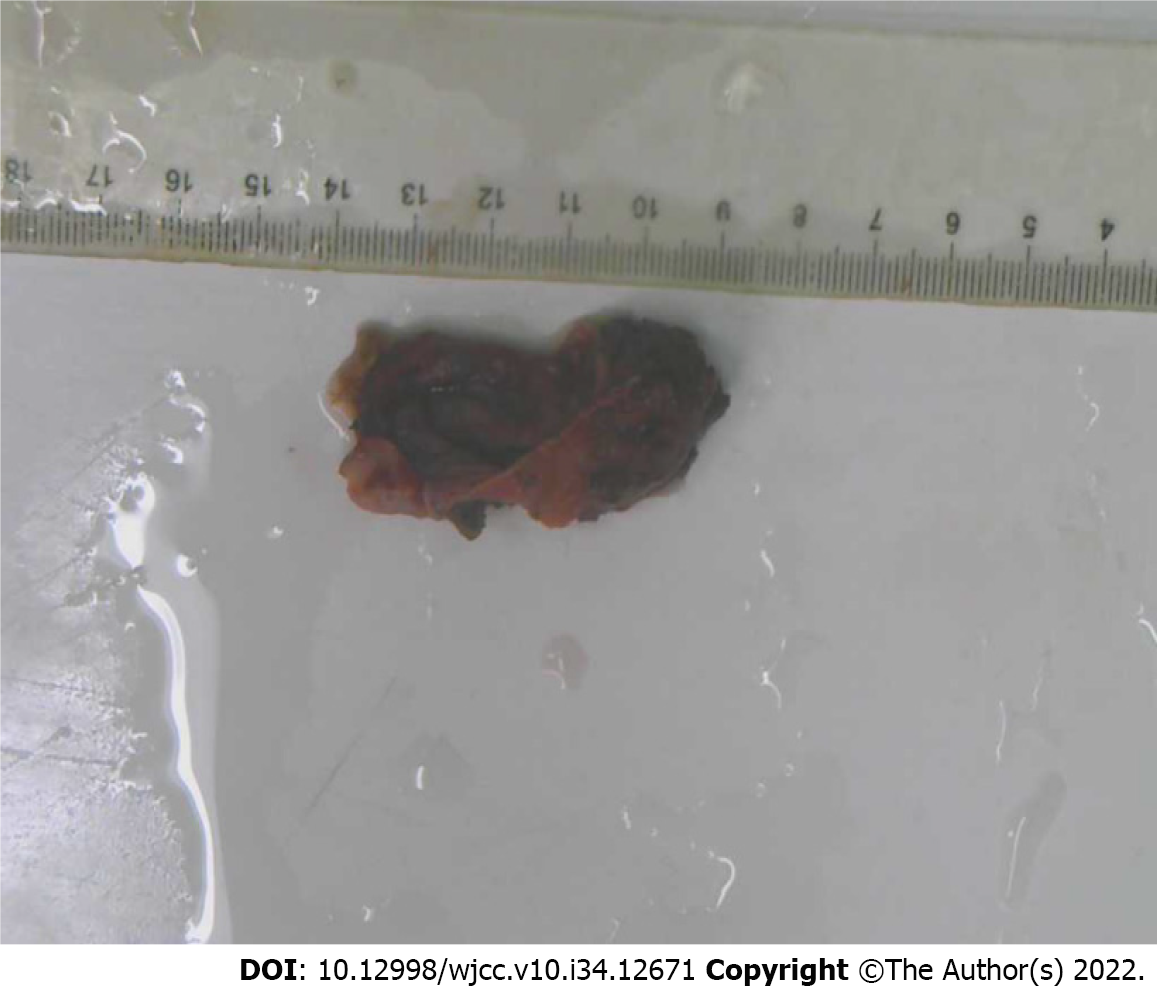
Figure 2 Resected section of the cystic mass postoperatively.
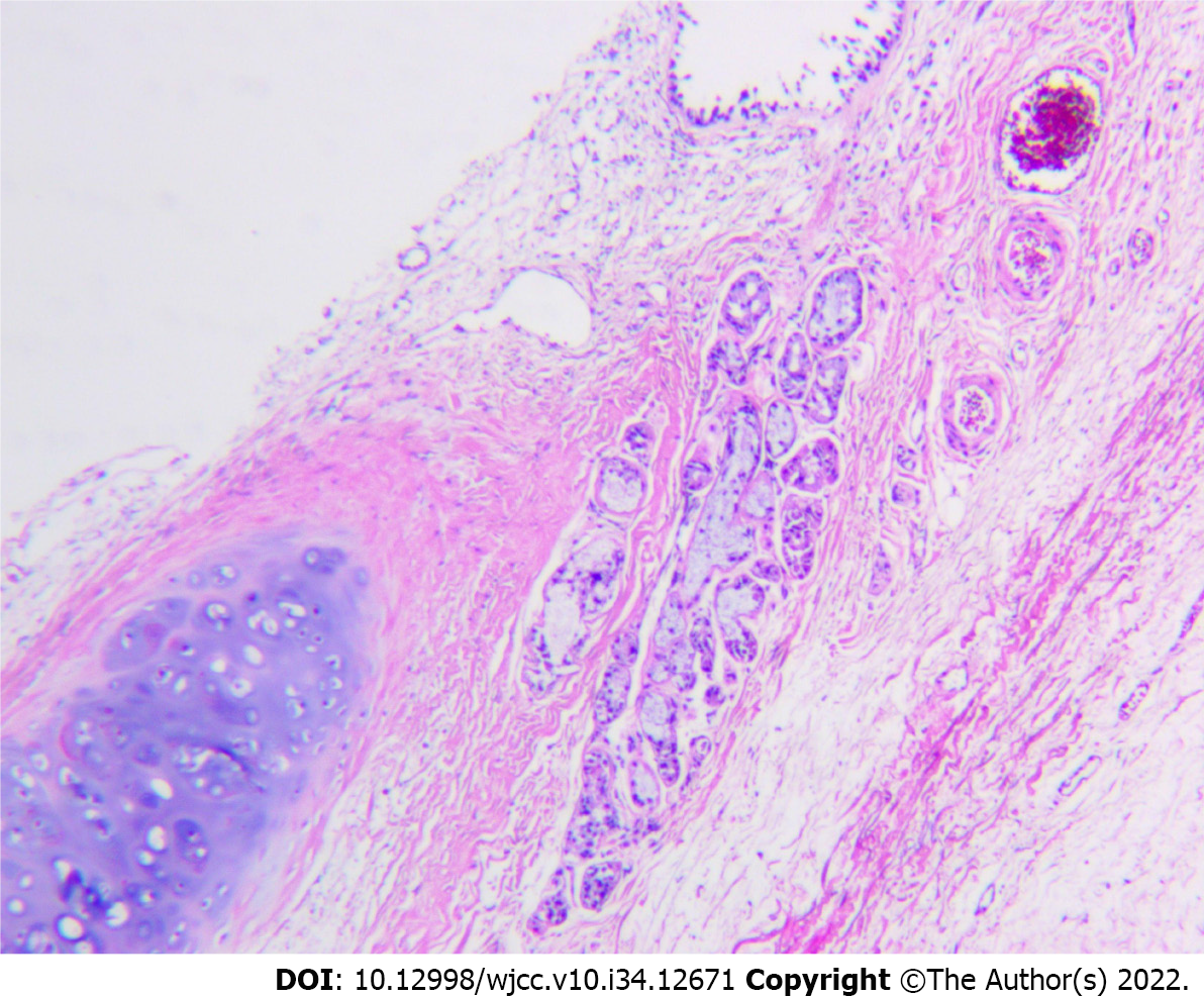
Figure 3 Cystic wall lined with columnar epithelium with cartilage and mucous glands.
Hematoxylin and eosin stain, 10× 4.
- Citation: Li C, Zhang XW, Zhao CA, Liu M. Abdominal bronchogenic cyst: A rare case report. World J Clin Cases 2022; 10(34): 12671-12677
- URL: https://www.wjgnet.com/2307-8960/full/v10/i34/12671.htm
- DOI: https://dx.doi.org/10.12998/wjcc.v10.i34.12671


