Copyright
©The Author(s) 2022.
World J Clin Cases. Dec 6, 2022; 10(34): 12631-12636
Published online Dec 6, 2022. doi: 10.12998/wjcc.v10.i34.12631
Published online Dec 6, 2022. doi: 10.12998/wjcc.v10.i34.12631
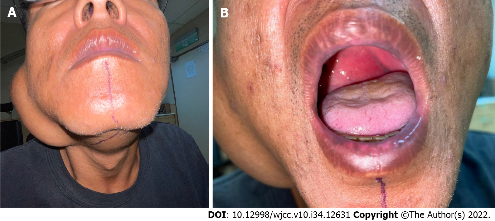
Figure 1 Parotid tumor images.
A: Outward appearance of the parotid tumor; B: As the patient opened his mouth, we can see the tumor that invaded into the oropharynx.
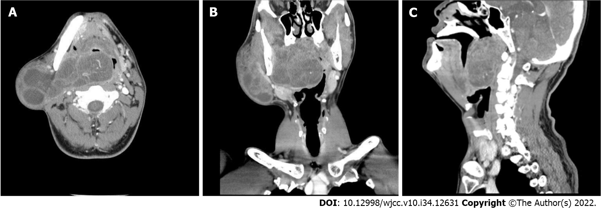
Figure 2 Computed tomography of the head and neck.
A: Axial computed tomography image shows a huge mass from the right parotid region with a deep extension through the lateral pharyngeal region to the retropharyngeal region; B: Coronal computed tomography image shows that the tumor occupied most of the space of the oropharynx; C: Sagittal computed tomography image shows obliteration of the nasopharynx to oropharynx.
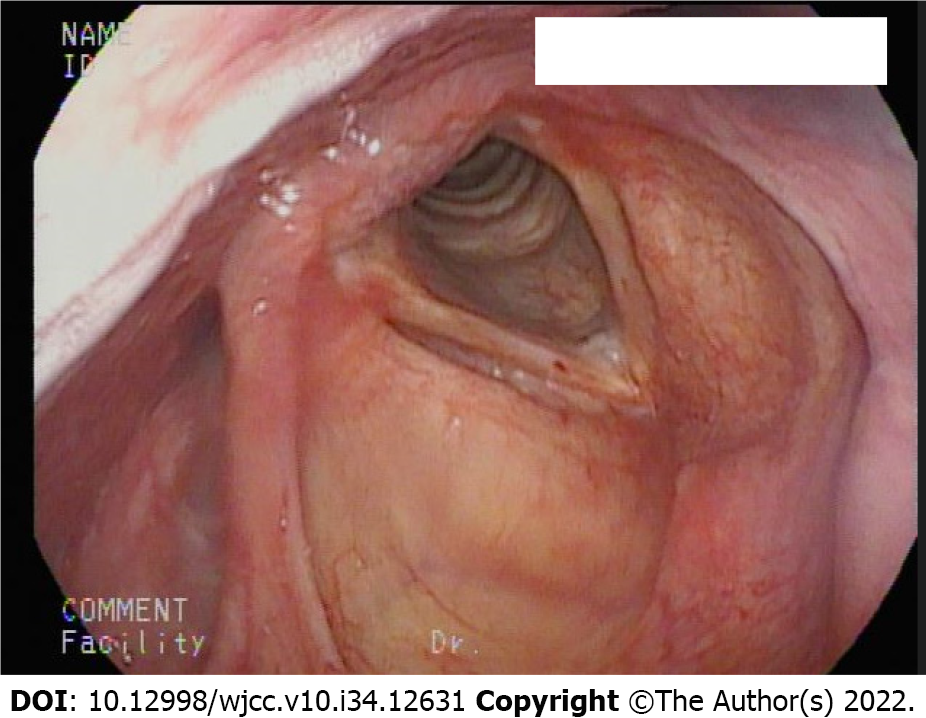
Figure 3 Fiberoptic examinaiton via an oral approach demonstrated clear view of the laryngeal inlet after bypassing the tumor.
- Citation: Lin TC, Lai YW, Wu SH. Emergent use of tube tip in pharynx technique in “cannot intubate cannot oxygenate” situation: A case report. World J Clin Cases 2022; 10(34): 12631-12636
- URL: https://www.wjgnet.com/2307-8960/full/v10/i34/12631.htm
- DOI: https://dx.doi.org/10.12998/wjcc.v10.i34.12631


