Copyright
©The Author(s) 2022.
World J Clin Cases. Dec 6, 2022; 10(34): 12610-12616
Published online Dec 6, 2022. doi: 10.12998/wjcc.v10.i34.12610
Published online Dec 6, 2022. doi: 10.12998/wjcc.v10.i34.12610
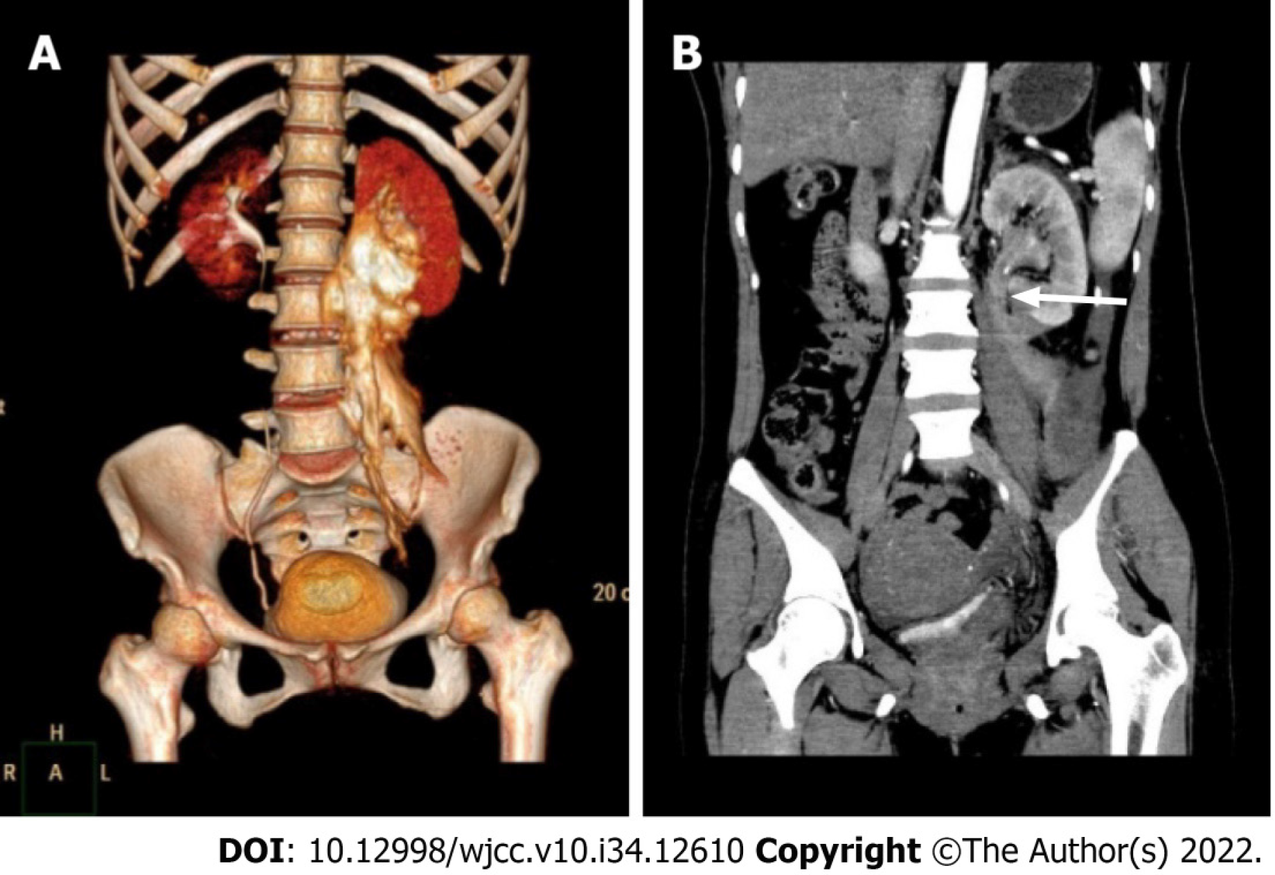
Figure 1 Preoperative computed tomography.
A: Computed tomography (CT) urography three-dimensional reconstruction showed retroperitoneal contrast extravasation with the most severe site in the ureteropelvic junction (UPJ) region; B: Enhanced CT showed left ureteral disruption just below the UPJ (white arrow).
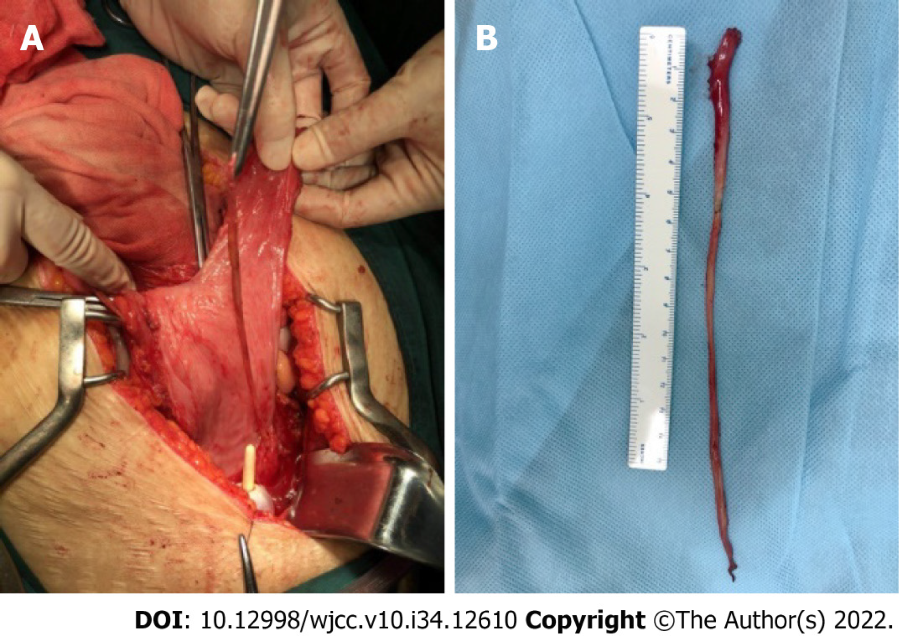
Figure 2 The completely avulsed ureter.
A: A long isolated devitalized ureter was noticed within the bladder intraoperatively; B: The full-length avulsed ureter in vitro.
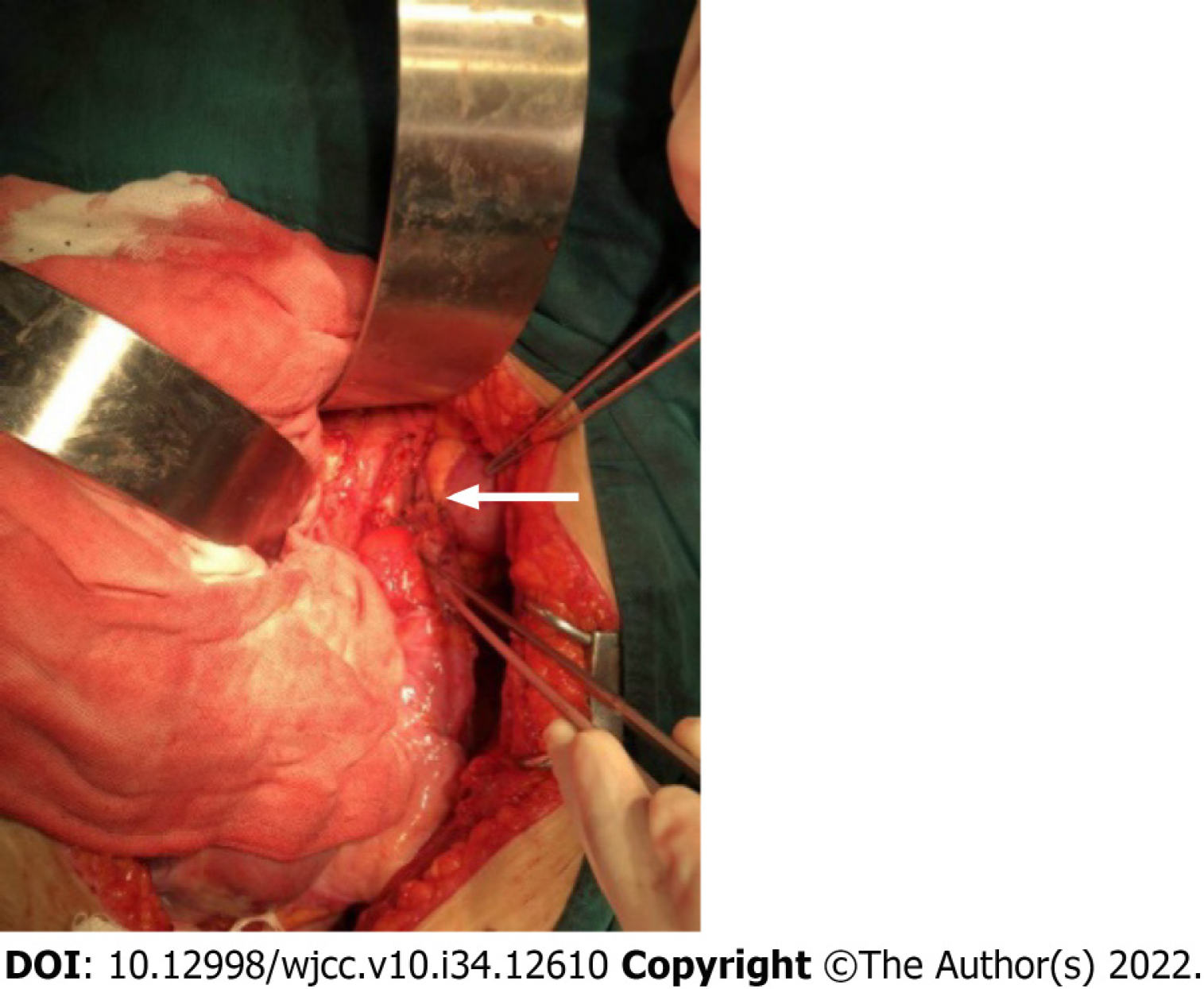
Figure 3 Ureteral reconstruction with an extended Boari flap intraoperatively.
The white arrow shows a tension-free anastomosis.
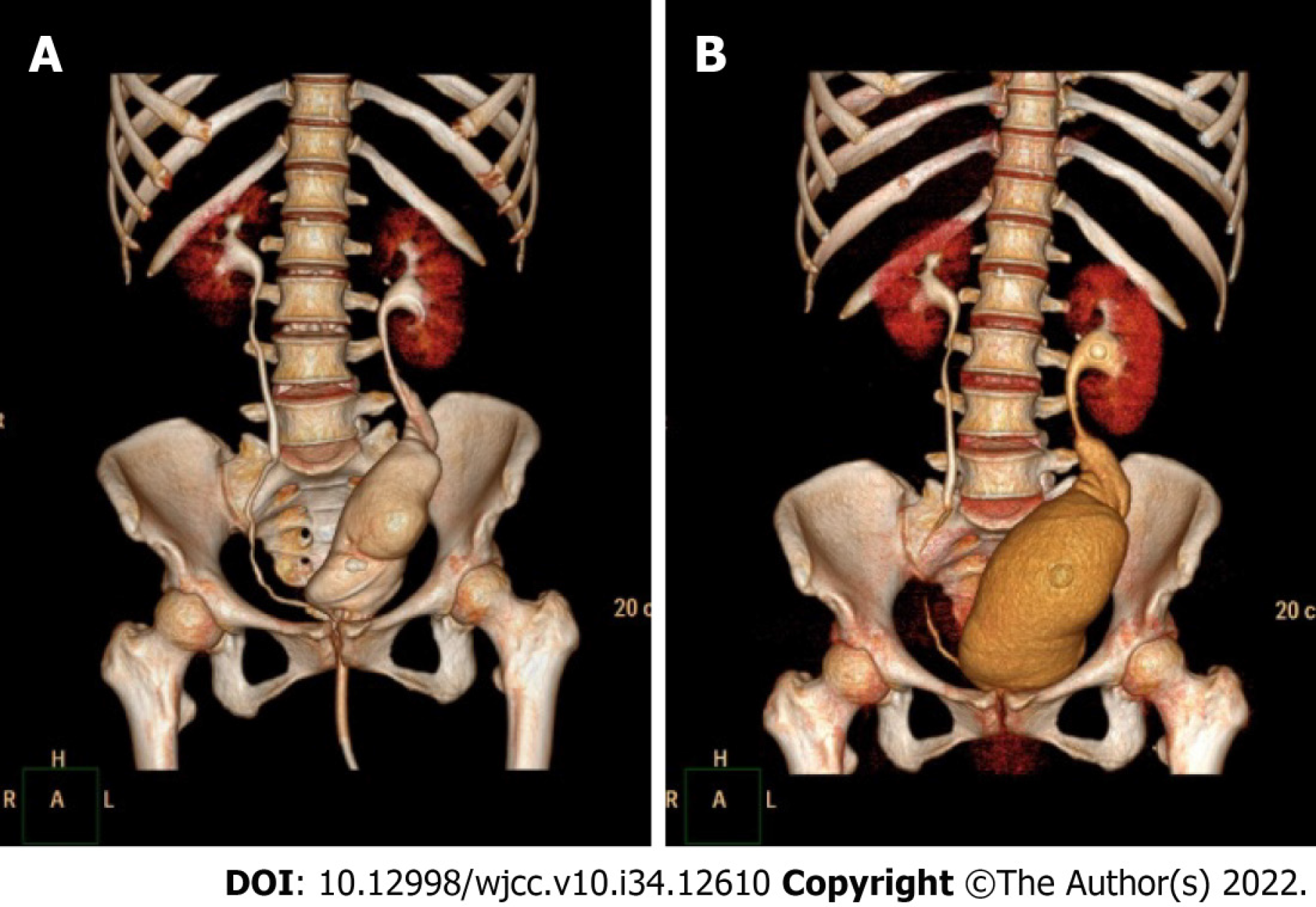
Figure 4 Postoperative computed tomography urography.
A and B: Computed tomography urography three-dimensional reconstruction at 14 d (A) and 2 mo postoperatively. Note that bladder volume is visibly larger at 2 mo postoperatively.
- Citation: Zhong MZ, Huang WN, Huang GX, Zhang EP, Gan L. Long-term results of extended Boari flap technique for management of complete ureteral avulsion: A case report. World J Clin Cases 2022; 10(34): 12610-12616
- URL: https://www.wjgnet.com/2307-8960/full/v10/i34/12610.htm
- DOI: https://dx.doi.org/10.12998/wjcc.v10.i34.12610


