Copyright
©The Author(s) 2022.
World J Clin Cases. Nov 26, 2022; 10(33): 12440-12446
Published online Nov 26, 2022. doi: 10.12998/wjcc.v10.i33.12440
Published online Nov 26, 2022. doi: 10.12998/wjcc.v10.i33.12440
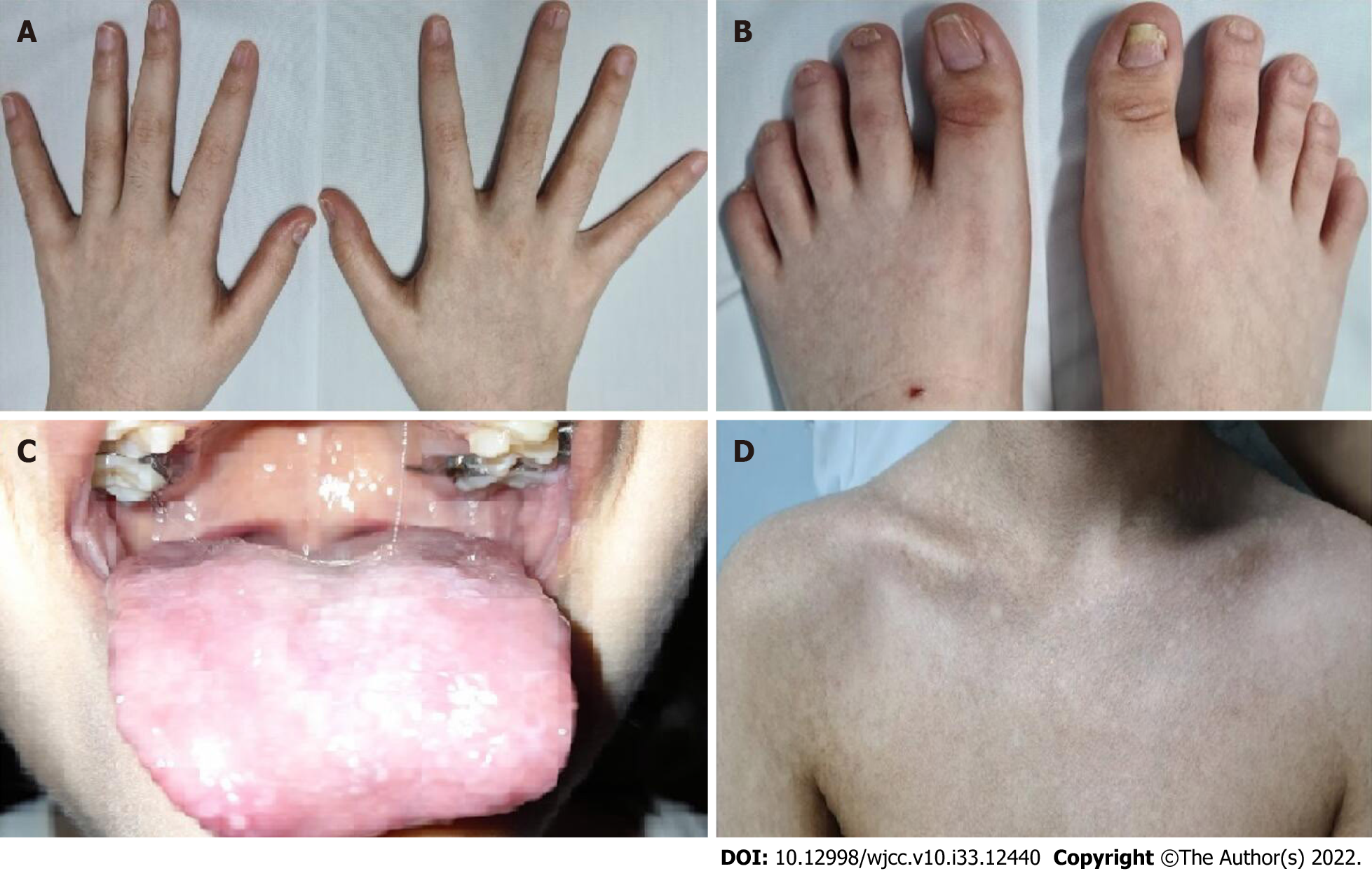
Figure 1 Clinical findings in the patient.
A: Dystrophic fingernails; B: Dystrophic toenails; C: White patches on the tongue representing leukoplakia; D: Upper trunk showing reticulate skin pigmentation.
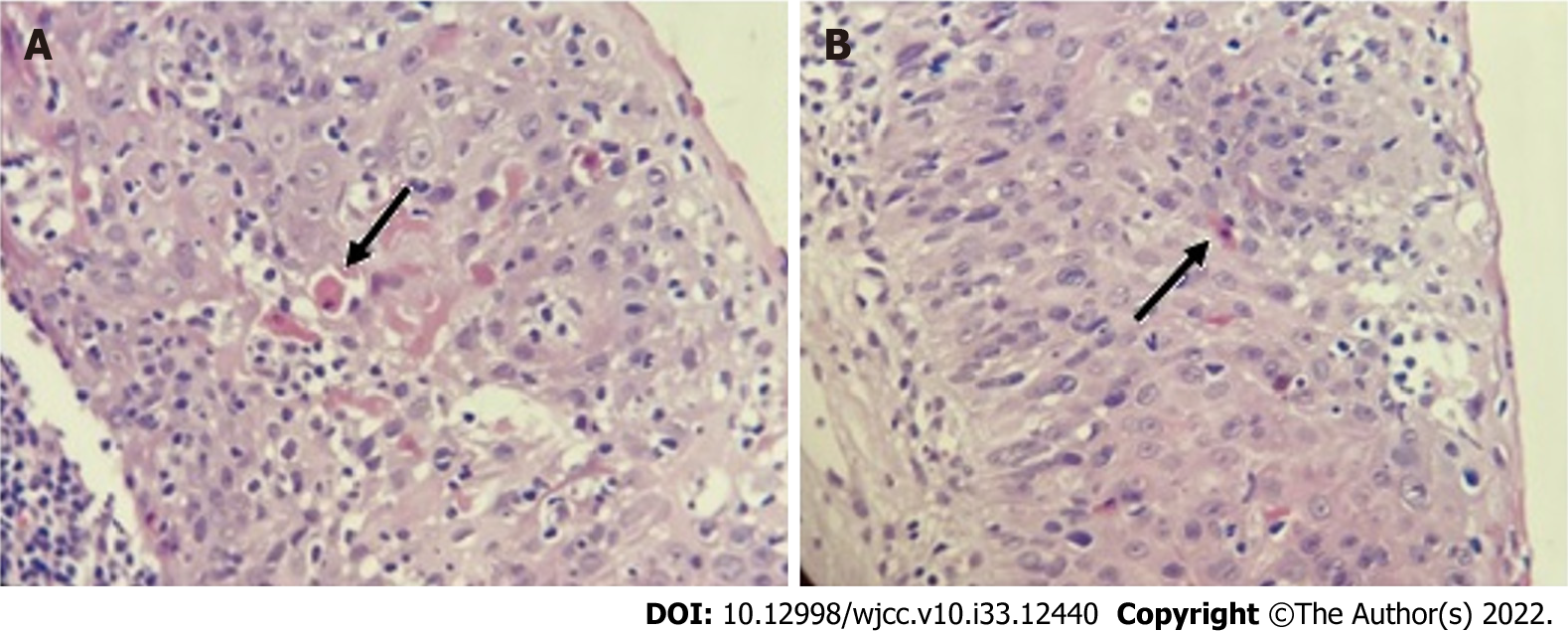
Figure 2 Histopathological study.
A and B: Skin biopsy images showing dyskeratocytes. The arrows point to these abnormal cells.
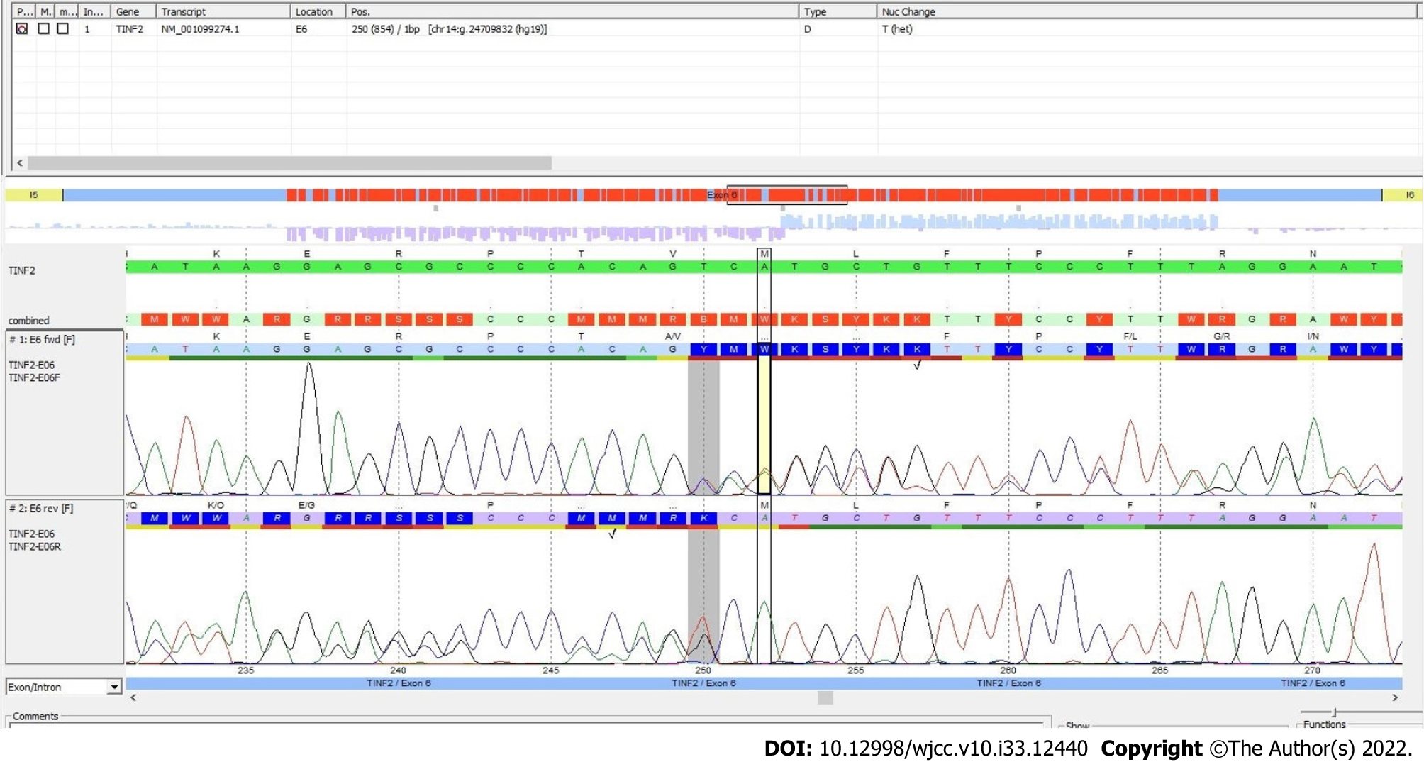
Figure 3 Sequencing study.
Plot showing the identification of the mutation in the TINF2 gene (deletion of the T in the position 250; shaded area). Courtesy of Centogene AG, Rostock, Germany.
- Citation: Picos-Cárdenas VJ, Beltrán-Ontiveros SA, Cruz-Ramos JA, Contreras-Gutiérrez JA, Arámbula-Meraz E, Angulo-Rojo C, Guadrón-Llanos AM, Leal-León EA, Cedano-Prieto DM, Meza-Espinoza JP. Novel TINF2 gene mutation in dyskeratosis congenita with extremely short telomeres: A case report. World J Clin Cases 2022; 10(33): 12440-12446
- URL: https://www.wjgnet.com/2307-8960/full/v10/i33/12440.htm
- DOI: https://dx.doi.org/10.12998/wjcc.v10.i33.12440


