Copyright
©The Author(s) 2022.
World J Clin Cases. Nov 26, 2022; 10(33): 12328-12336
Published online Nov 26, 2022. doi: 10.12998/wjcc.v10.i33.12328
Published online Nov 26, 2022. doi: 10.12998/wjcc.v10.i33.12328
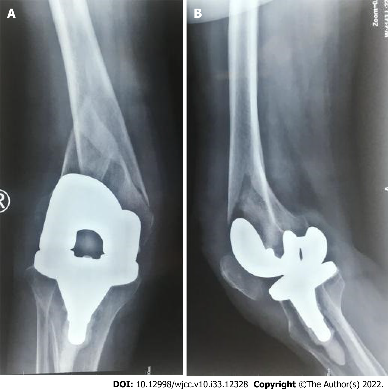
Figure 1 Preoperative images of case 1.
A: Anteroposterior radiographs of the right distal femur at the time of presentation showing the supracondylar femoral fracture above total knee arthroplasty which was non-comminuted and oblique; B: Lateral radiograph of the right periprosthetic supracondylar femoral fracture.
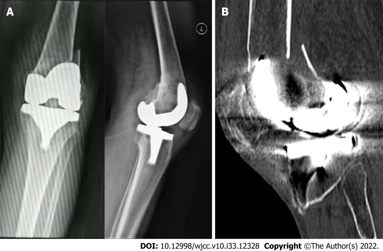
Figure 2 Preoperative images of case 2.
A: Plain radiographs of the left distal femur at the time of presentation showing the supracondylar femoral fracture just above total knee arthroplasty, which was non-comminuted and transverse; B: Sagittal computed tomography image of the left periprosthetic supracondylar femoral fracture.
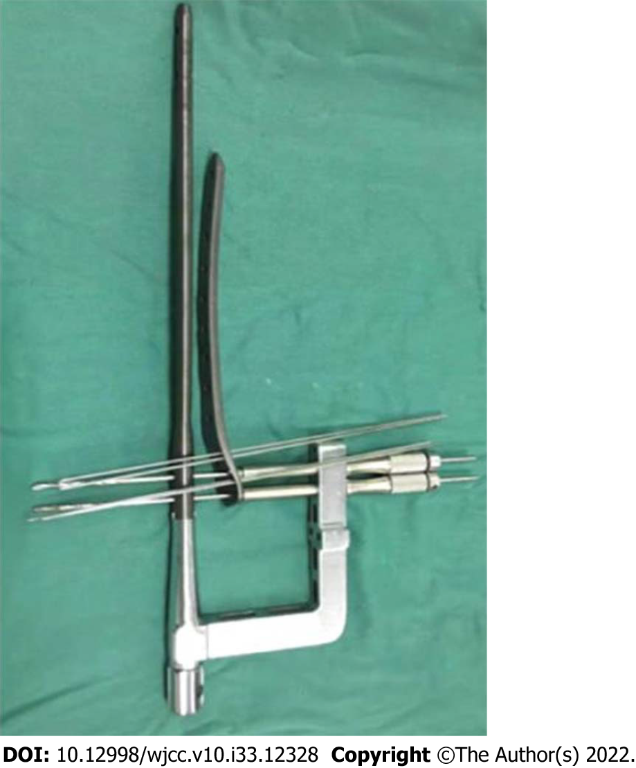
Figure 3 Implant of modified nail plate combination.
Image of implant showing a specially designed periprosthetic locking plate, a retrograde intramedullary nail, an intramedullary targeting arm, locking screw guide and k wires.
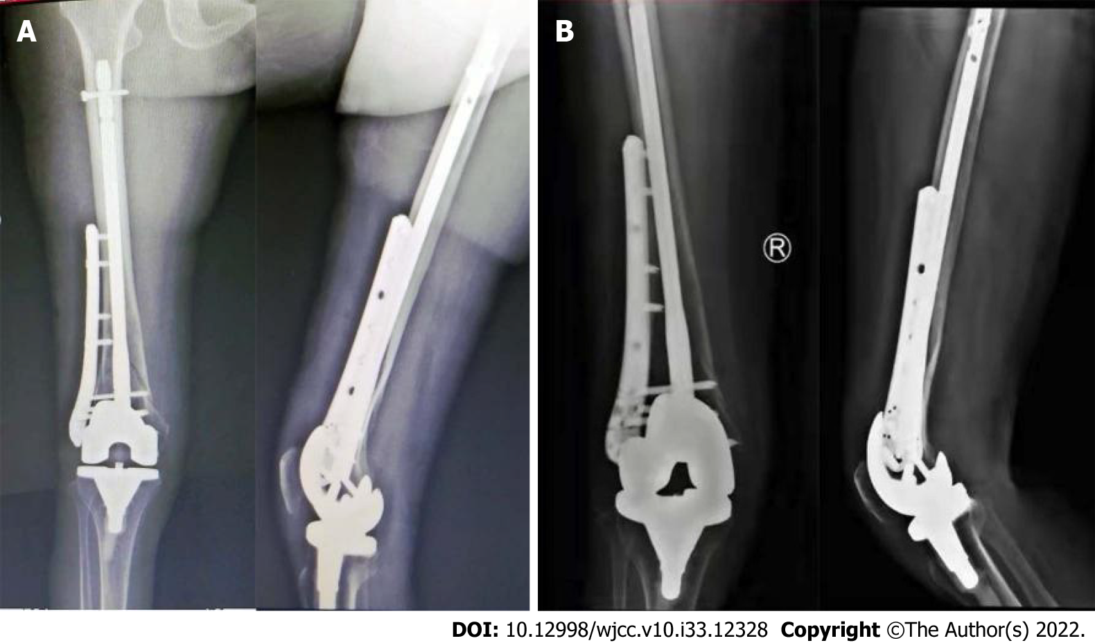
Figure 4 Postoperative and follow-up images of case 1.
A: Anteroposterior and lateral X-ray showing good reduction of periprosthetic supracondylar femoral fracture through modified nail plate combination; B: Anteroposterior and lateral X-ray taken 6 mo later showing evidence of union.
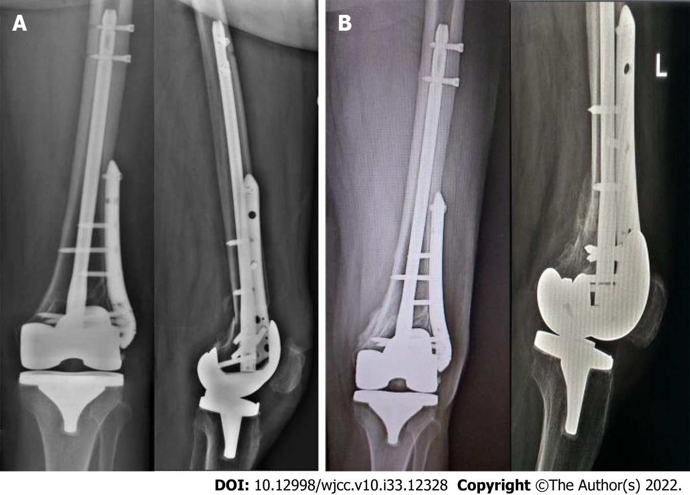
Figure 5 Postoperative and follow-up images of case 2.
A: Anteroposterior and lateral X-ray showing good reduction of periprosthetic supracondylar femoral fracture through modified nail plate combination; B: Anteroposterior and lateral X-ray taken 6 mo later showing evidence of union.
- Citation: Li QW, Wu B, Chen B. Modified fixation for periprosthetic supracondylar femur fractures: Two case reports and review of the literature. World J Clin Cases 2022; 10(33): 12328-12336
- URL: https://www.wjgnet.com/2307-8960/full/v10/i33/12328.htm
- DOI: https://dx.doi.org/10.12998/wjcc.v10.i33.12328


